Enterría-Morales Daniel, Del Rey Natalia López-González, Blesa Javier, López-López Ivette, Gallet Sarah, Prévot Vincent, López-Barneo José, d'Anglemont de Tassigny Xavier
Instituto de Biomedicina de Sevilla (IBiS), Hospital Universitario Virgen del Rocío, CSIC, Universidad de Sevilla, Seville, Spain.
Departamento de Fisiología Médica y Biofísica, Facultad de Medicina, Universidad de Sevilla, Seville, Spain.
Brain Commun. 2020 Aug 27;2(2):fcaa105. doi: 10.1093/braincomms/fcaa105. eCollection 2020.
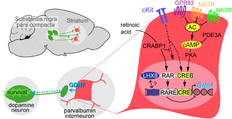
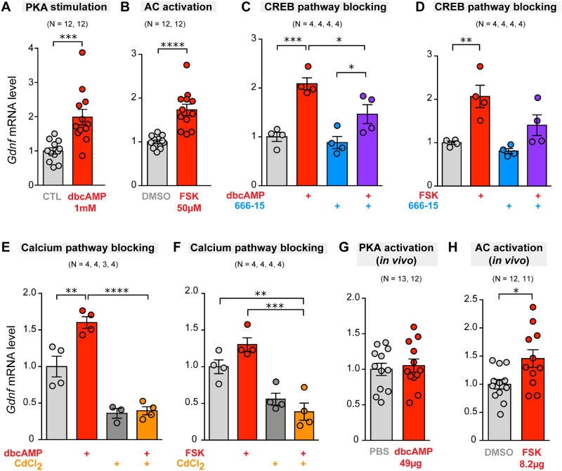
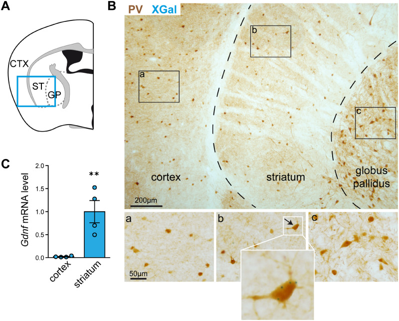
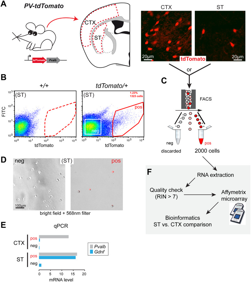
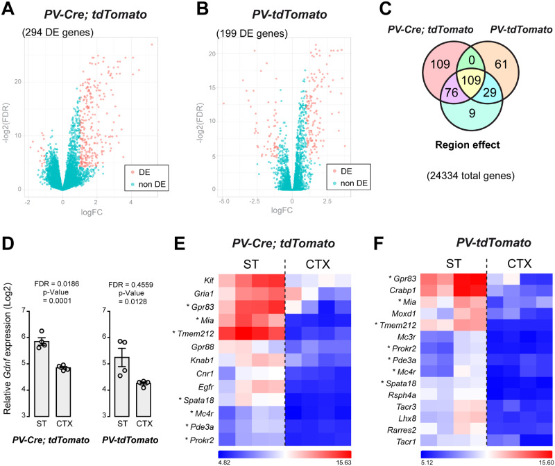
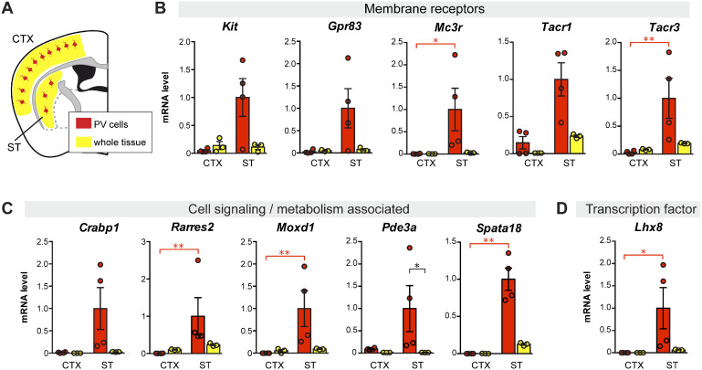
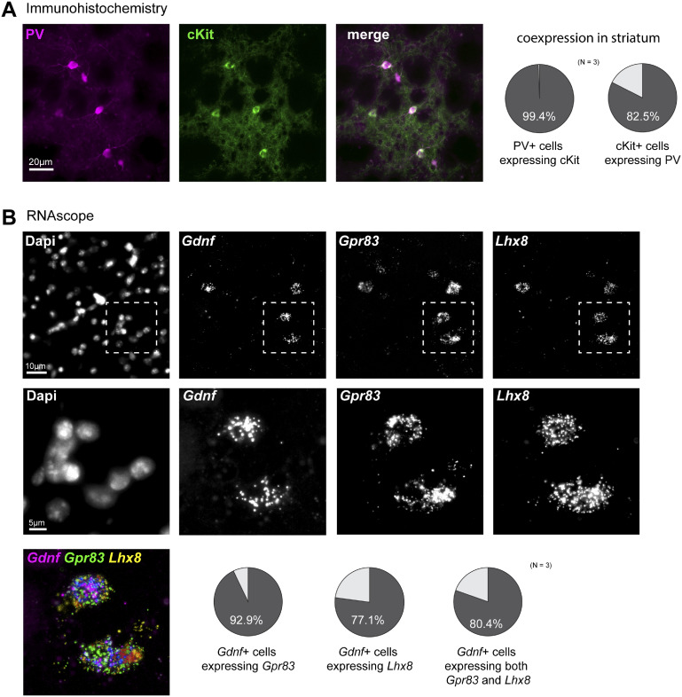
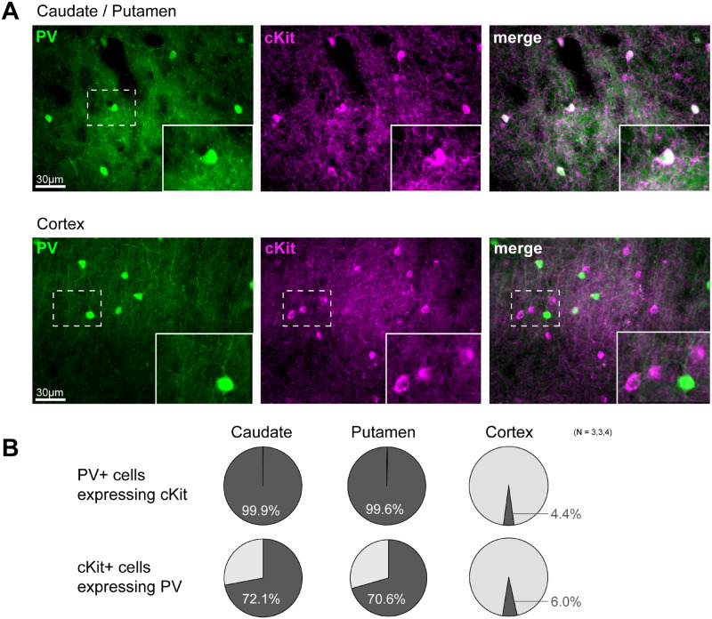
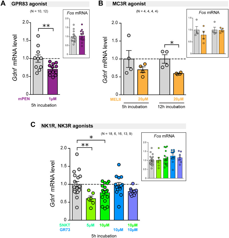
Administration of recombinant glial cell line-derived neurotrophic factor into the putamen has been tested in preclinical and clinical studies to evaluate its neuroprotective effects on the progressive dopaminergic neuronal degeneration that characterizes Parkinson's disease. However, intracerebral glial cell line-derived neurotrophic factor infusion is a challenging therapeutic strategy, with numerous potential technical and medical limitations. Most of these limitations could be avoided if the production of endogenous glial cell line-derived neurotrophic factor could be increased. Glial cell line-derived neurotrophic factor is naturally produced in the striatum from where it exerts a trophic action on the nigrostriatal dopaminergic pathway. Most of striatal glial cell line-derived neurotrophic factor is synthesized by a subset of GABAergic interneurons characterized by the expression of parvalbumin. We sought to identify molecular targets specific to those neurons and which are putatively associated with glial cell line-derived neurotrophic factor synthesis. To this end, the transcriptomic differences between glial cell line-derived neurotrophic factor-positive parvalbumin neurons in the striatum and parvalbumin neurons located in the nearby cortex, which do not express glial cell line-derived neurotrophic factor, were analysed. Using mouse reporter models, we have defined the genomic signature of striatal parvalbumin interneurons obtained by fluorescence-activated cell sorting followed by microarray comparison. Short-listed genes were validated by additional histological and molecular analyses. These genes code for membrane receptors (, , , , ), cytosolic proteins (, , , ) and a transcription factor (). We also found the proto-oncogene cKit to be highly specific of parvalbumin interneurons in the non-human primate striatum, thus highlighting a conserved expression between species and suggesting that specific genes identified in mouse parvalbumin neurons could be putative targets in the human brain. Pharmacological stimulation of four G-protein-coupled receptors enriched in the striatal parvalbumin interneurons inhibited expression presumably by decreasing cyclic adenosine monophosphate formation. Additional experiments with pharmacological modulators of adenylyl cyclase and protein kinase A indicated that this pathway is a relevant intracellular route to induce gene activation. This preclinical study is an important step in the ongoing development of a specific pro-endo-glial cell line-derived neurotrophic factor pharmacological strategy to treat Parkinson's disease.
在临床前和临床研究中,已对向壳核注射重组胶质细胞源性神经营养因子进行了测试,以评估其对帕金森病特征性的进行性多巴胺能神经元变性的神经保护作用。然而,脑内注射胶质细胞源性神经营养因子是一种具有挑战性的治疗策略,存在许多潜在的技术和医学限制。如果能够增加内源性胶质细胞源性神经营养因子的产生,这些限制中的大多数是可以避免的。胶质细胞源性神经营养因子在纹状体中天然产生,它在那里对黑质纹状体多巴胺能通路发挥营养作用。纹状体中的大多数胶质细胞源性神经营养因子是由一小部分以表达小白蛋白为特征的γ-氨基丁酸能中间神经元合成的。我们试图确定这些神经元特有的分子靶点,这些靶点可能与胶质细胞源性神经营养因子的合成有关。为此,分析了纹状体中胶质细胞源性神经营养因子阳性小白蛋白神经元与位于附近皮质但不表达胶质细胞源性神经营养因子的小白蛋白神经元之间的转录组差异。使用小鼠报告模型,我们通过荧光激活细胞分选然后进行微阵列比较,确定了纹状体小白蛋白中间神经元的基因组特征。通过额外的组织学和分子分析对入围基因进行了验证。这些基因编码膜受体(、、、、)、胞质蛋白(、、、)和一种转录因子()。我们还发现原癌基因cKit在非人类灵长类动物纹状体的小白蛋白中间神经元中具有高度特异性,从而突出了物种间的保守表达,并表明在小鼠小白蛋白神经元中鉴定出的特定基因可能是人类大脑中的潜在靶点。对富含纹状体小白蛋白中间神经元的四种G蛋白偶联受体进行药理刺激,可能通过减少环磷酸腺苷的形成来抑制表达。使用腺苷酸环化酶和蛋白激酶A的药理调节剂进行的额外实验表明,该途径是诱导基因激活的一条相关细胞内途径。这项临床前研究是正在进行的开发治疗帕金森病的特异性促内源性胶质细胞源性神经营养因子药理策略的重要一步。