Laboratoire de Biochimie Médicale et Biologie Moléculaire, Université de Reims Champagne-Ardenne, 51097 Reims, France.
CNRS UMR 7369, Matrice Extracellulaire et Dynamique Cellulaire-MEDyC, 51097 Reims, France.
Molecules. 2020 Sep 19;25(18):4300. doi: 10.3390/molecules25184300.
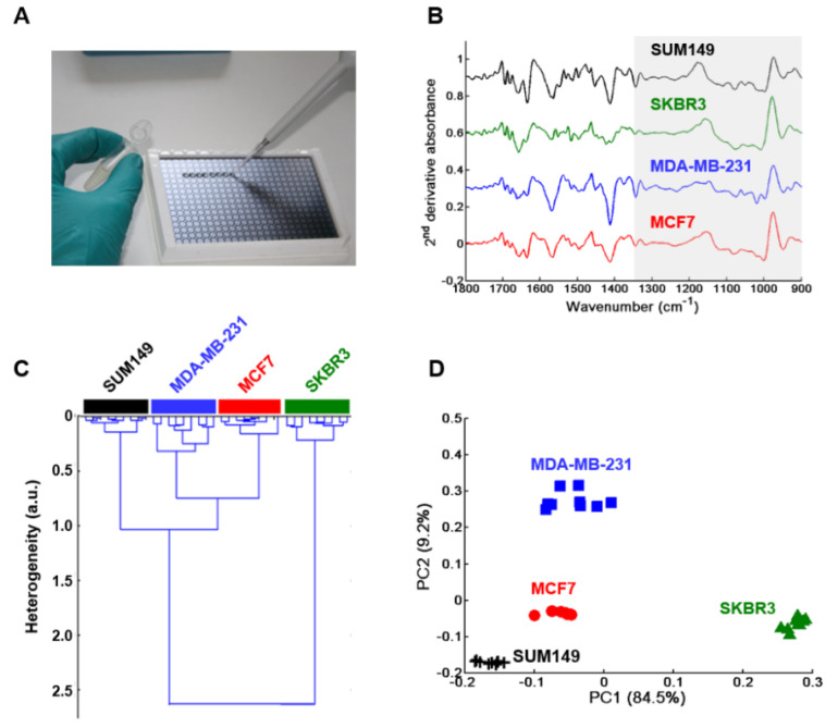
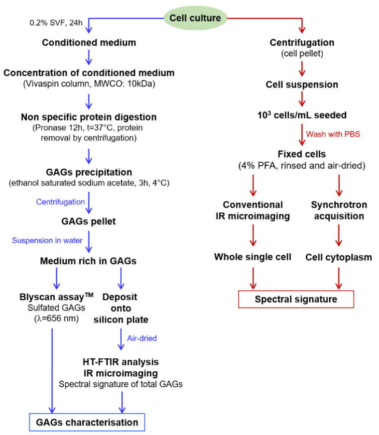
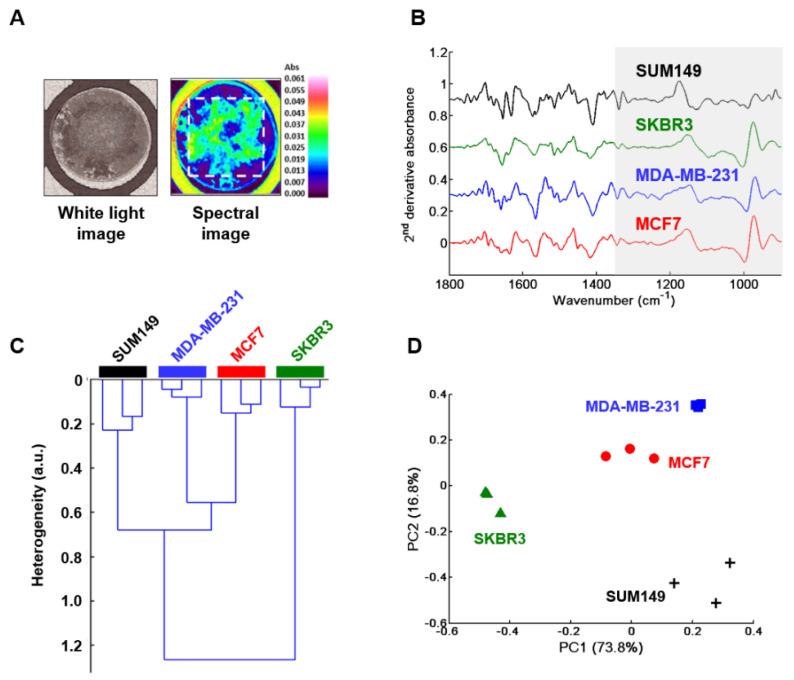
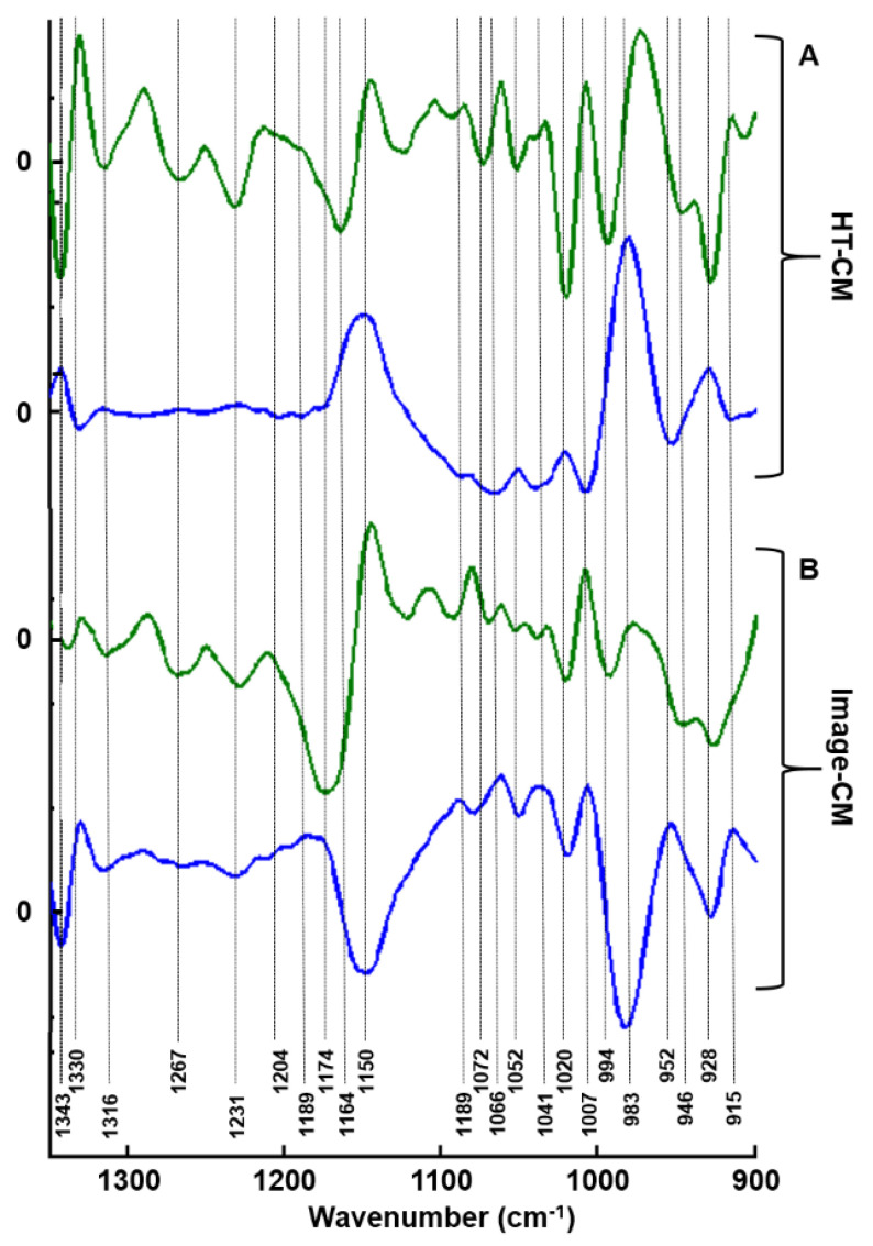
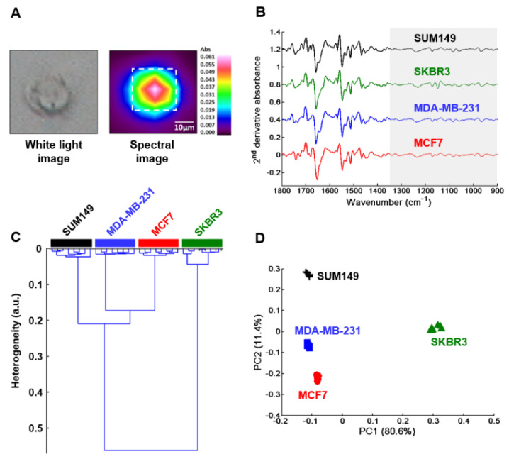
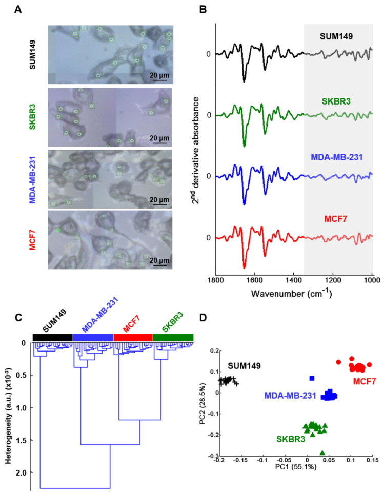
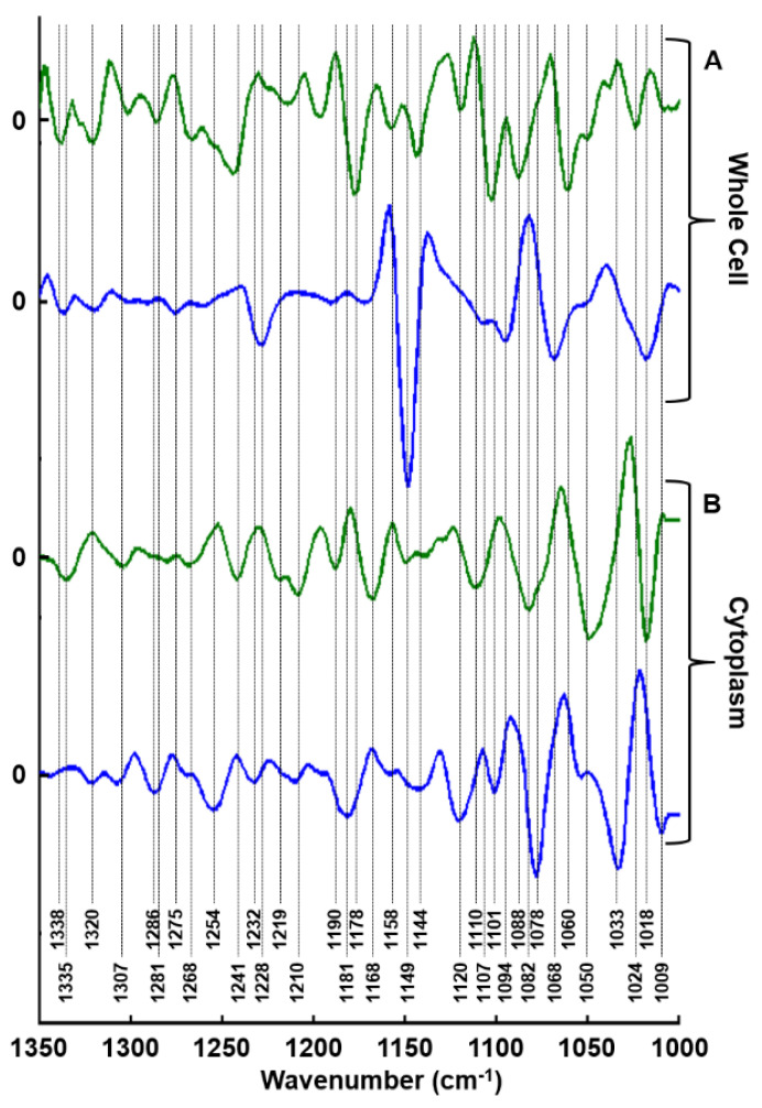
Glycosaminoglycans (GAGs)/proteoglycans (PGs) play a pivotal role in the metastasis of inflammatory breast cancer (IBC). They represent biomarkers and targets in diagnosis and treatment of different cancers including breast cancer. Thus, GAGs/PGs could represent potential prognostic/diagnostic biomarkers for IBC. In the present study, non-IBC MDA-MB-231, MCF7, SKBR3 cells and IBC SUM149 cells, as well as their GAG secretome were analyzed. The latter was measured in toto as dried drops with high-throughput (HT) Fourier Transform InfraRed (FTIR) spectroscopy and imaging. FTIR imaging was also employed to investigate single whole breast cancer cells while synchrotron-FTIR microspectroscopy was used to specifically target their cytoplasms. Data were analyzed by hierarchical cluster analysis and principal components analysis. Results obtained from HT-FTIR analysis of GAG drops showed that the inter-group variability enabled us to delineate between cell types in the GAG absorption range 1350-800 cm. Similar results were obtained for FTIR imaging of GAG extracts and fixed single whole cells. Synchrotron-FTIR data from cytoplasms allowed discrimination between non-IBC and IBC. Thus, by using GAG specific region, not only different breast cancer cell lines could be differentiated, but also non-IBC from IBC cells. This could be a potential diagnostic spectral marker for IBC detection useful for patient management.
糖胺聚糖(GAGs)/蛋白聚糖(PGs)在炎症性乳腺癌(IBC)的转移中起着关键作用。它们是不同癌症(包括乳腺癌)诊断和治疗的生物标志物和靶点。因此,GAGs/PGs 可能是 IBC 的潜在预后/诊断生物标志物。在本研究中,分析了非 IBC MDA-MB-231、MCF7、SKBR3 细胞和 IBC SUM149 细胞及其 GAG 分泌组。后者通过高通量(HT)傅里叶变换红外(FTIR)光谱和成像以干燥液滴的形式进行了全面测量。FTIR 成像还用于研究单个全乳腺癌细胞,而同步辐射-FTIR 微光谱则用于专门针对其细胞质。通过层次聚类分析和主成分分析对数据进行了分析。通过对 GAG 液滴的 HT-FTIR 分析获得的数据表明,组间变异性使我们能够在 GAG 吸收范围 1350-800 cm 内区分细胞类型。GAG 提取物和固定的单个全细胞的 FTIR 成像也获得了类似的结果。来自细胞质的同步辐射-FTIR 数据允许区分非 IBC 和 IBC。因此,通过使用 GAG 特定区域,不仅可以区分不同的乳腺癌细胞系,还可以区分非 IBC 和 IBC 细胞。这可能是一种潜在的用于 IBC 检测的诊断光谱标记物,有助于患者管理。