Key Laboratory for Biomedical Engineering of Ministry of Education, College of Biomedical Engineering & Instrument Science, Zhejiang University, Hangzhou, Zhejiang, China.
Radiology, Beth Israel Deaconess Medical Center and Harvard Medical School, Boston, MA, USA.
Fluids Barriers CNS. 2020 Sep 22;17(1):58. doi: 10.1186/s12987-020-00218-z.
The choroid plexus is a major contributor to the generation of cerebrospinal fluid (CSF) and the maintenance of its electrolyte and metabolite balance. Here, we sought to characterize the blood flow dynamics of the choroid plexus using arterial spin labeling (ASL) MRI to establish ASL as a non-invasive tool for choroid plexus function and disease studies.
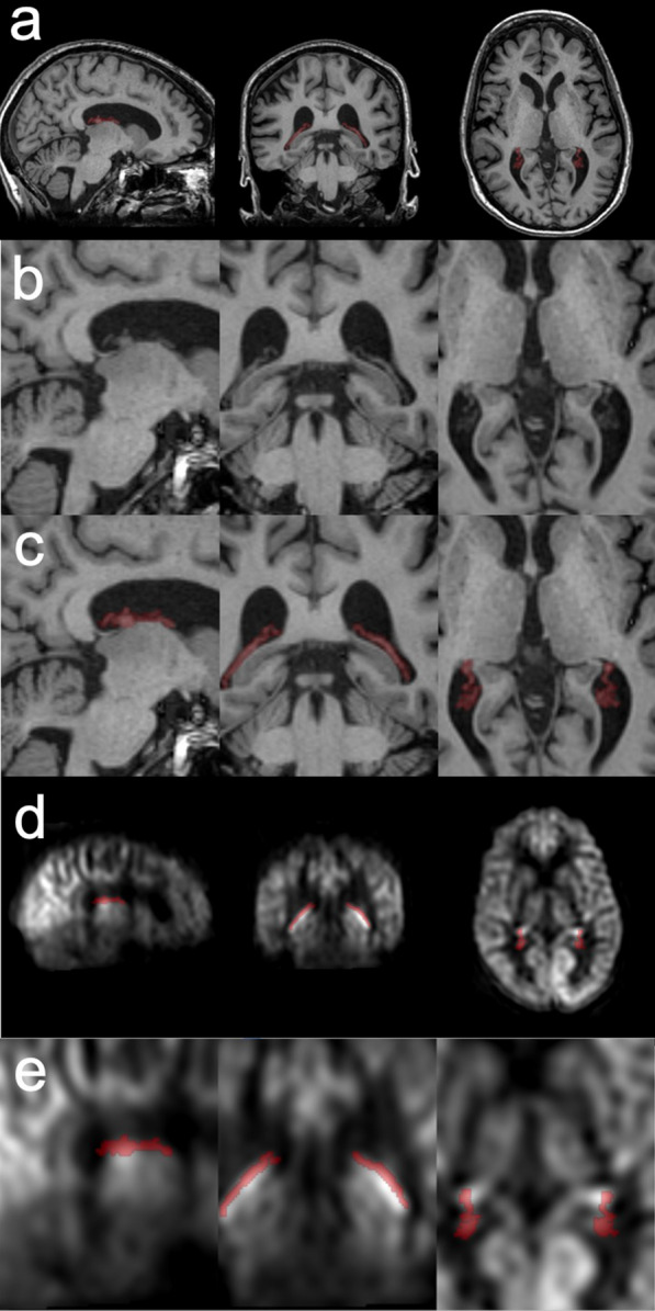
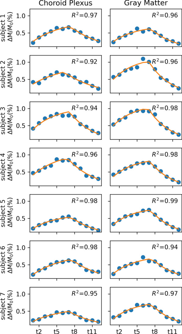
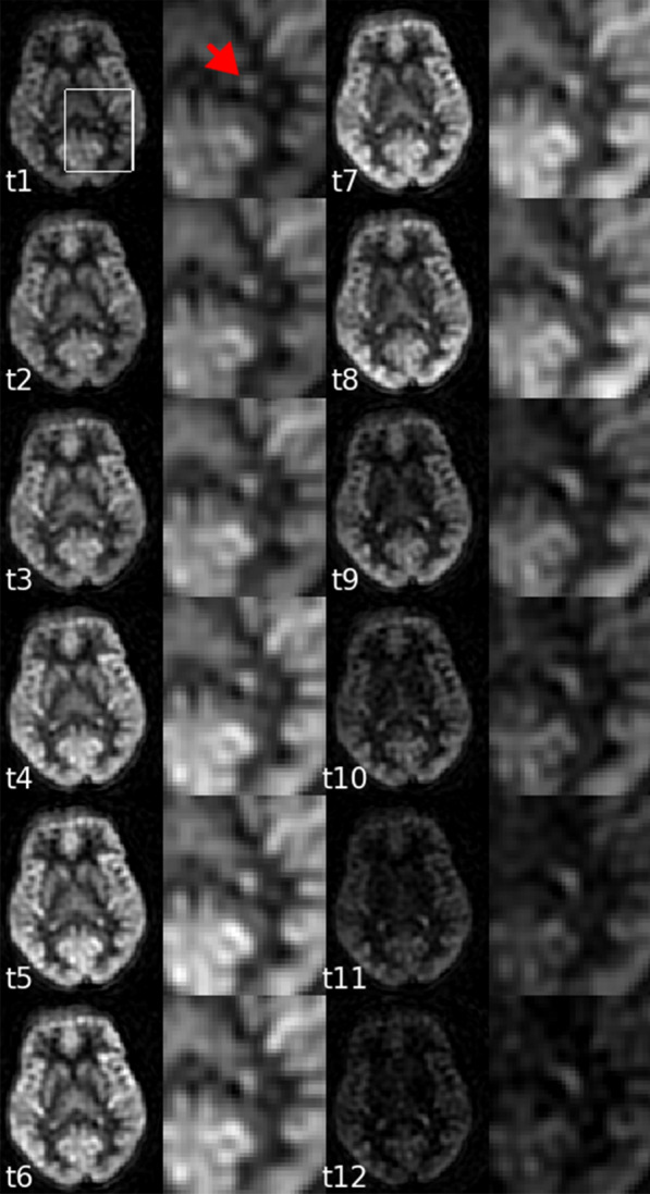
Seven healthy volunteers were imaged on a 3T MR scanner. ASL images were acquired with 12 labeling durations and post labeling delays. Regions of the choroid plexus were manually segmented on high-resolution T weighted images. Choroid plexus perfusion was characterized with a dynamic ASL perfusion model. Cerebral gray matter perfusion was also quantified for comparison.
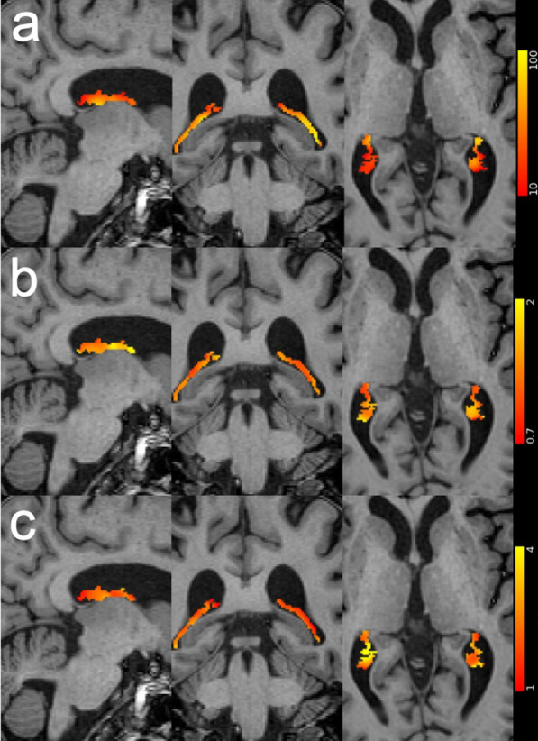
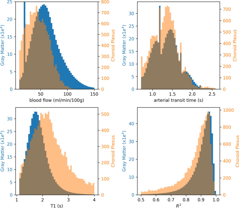
Kinetics of the ASL signal were clearly different in the choroid plexus than in gray matter. The choroid plexus has a significantly longer T than the gray matter (2.33 ± 0.30 s vs. 1.85 ± 0.10 s, p < 0.02). The arterial transit time was 1.24 ± 0.20 s at the choroid plexus. The apparent blood flow to the choroid plexus was measured to be 39.5 ± 10.1 ml/100 g/min and 0.80 ± 0.31 ml/min integrated over the posterior lateral ventricles in both hemispheres. Correction with the choroid plexus weight yielded a blood flow of 80 ml/100 g/min.
Our findings suggest that ASL can provide a clinically feasible option to quantify the dynamic characteristics of choroid plexus blood flow. It also provides useful reference values of the choroid plexus perfusion. The long T of the choroid plexus may suggest the transport of water from arterial blood to the CSF, potentially providing a method to quantify CSF generation.
脉络丛是产生脑脊液(CSF)和维持其电解质和代谢物平衡的主要贡献者。在这里,我们试图使用动脉自旋标记(ASL)MRI 来描述脉络丛的血流动力学,将 ASL 确立为一种用于脉络丛功能和疾病研究的非侵入性工具。
对 7 名健康志愿者在 3T MR 扫描仪上进行成像。使用 12 个标记持续时间和标记后延迟采集 ASL 图像。在高分辨率 T 加权图像上手动分割脉络丛区域。使用动态 ASL 灌注模型来描述脉络丛灌注。还定量了脑灰质灌注以进行比较。
ASL 信号的动力学在脉络丛和灰质之间明显不同。脉络丛的 T1 明显长于灰质(2.33±0.30 s 比 1.85±0.10 s,p<0.02)。脉络丛的动脉转运时间为 1.24±0.20 s。测量到脉络丛的表观血流为 39.5±10.1 ml/100 g/min,双侧后外侧脑室的积分值为 0.80±0.31 ml/min。用脉络丛重量校正后,血流为 80 ml/100 g/min。
我们的研究结果表明,ASL 可以提供一种可行的临床方法来量化脉络丛血流的动态特征。它还提供了脉络丛灌注的有用参考值。脉络丛的长 T1 可能表明水从动脉血向 CSF 的转运,这可能提供了一种量化 CSF 生成的方法。