Verheijen Nanne, Suttorp Christiaan M, van Rheden René E M, Regan Raymond F, Helmich Maria P A C, Kuijpers-Jagtman Anne Marie, Wagener Frank A D T G
Department of Dentistry - Orthodontics and Craniofacial Biology, Radboud University Medical Center, Nijmegen, Netherlands.
Radboud Institute for Molecular Life Sciences, Radboud University Medical Center, Nijmegen, Netherlands.
Front Cell Dev Biol. 2020 Aug 21;8:771. doi: 10.3389/fcell.2020.00771. eCollection 2020.
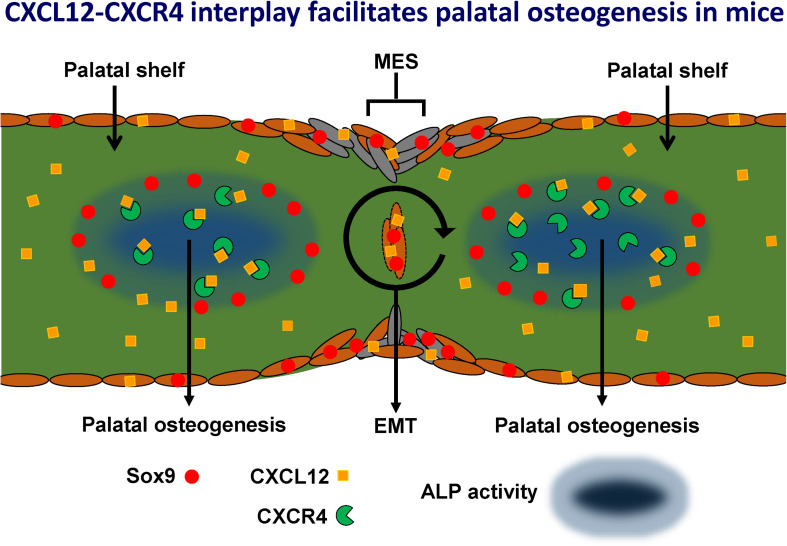
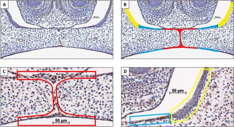
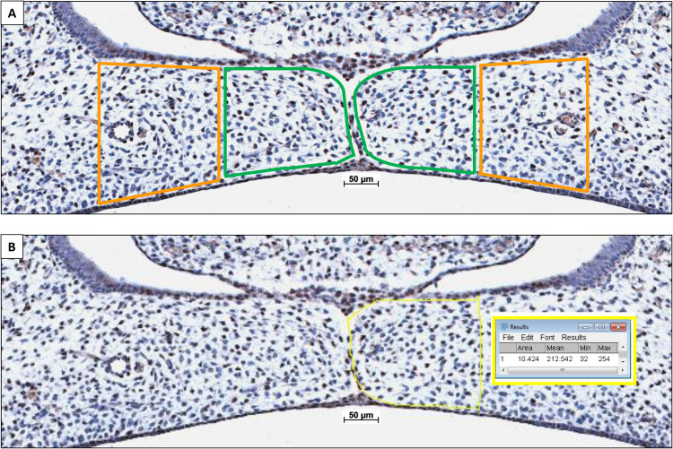
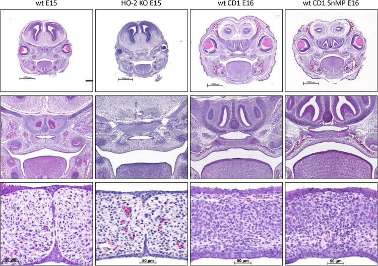
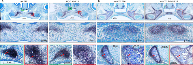
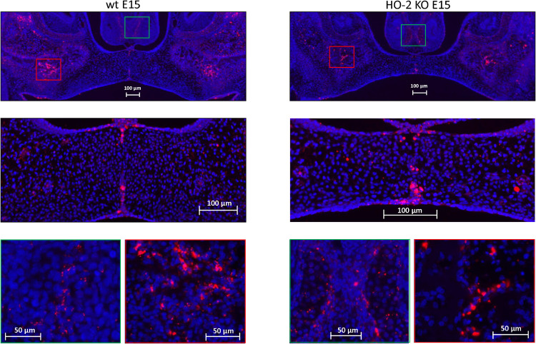
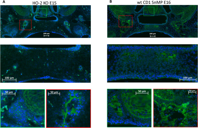
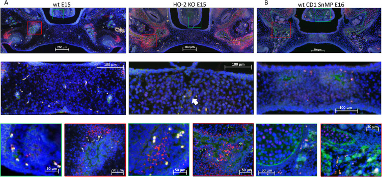
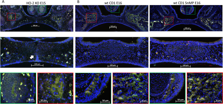
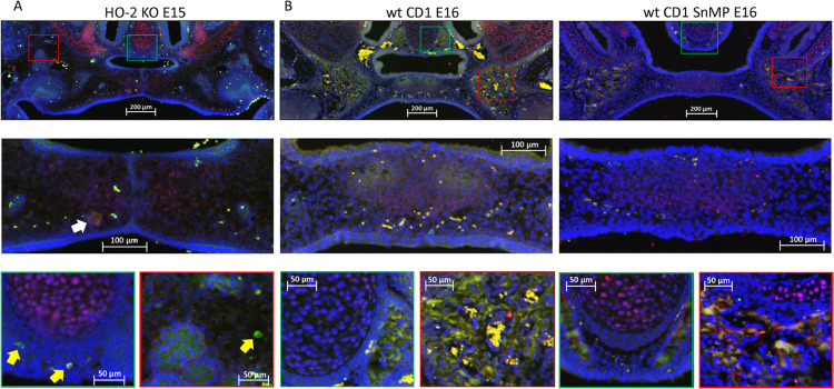
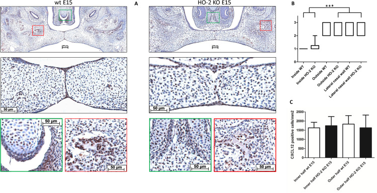
Cranial neural crest cells (CNCCs), identified by expression of transcription factor Sox9, migrate to the first branchial arch and undergo proliferation and differentiation to form the cartilage and bone structures of the orofacial region, including the palatal bone. Sox9 promotes osteogenic differentiation and stimulates CXCL12-CXCR4 chemokine-receptor signaling, which elevates alkaline phosphatase (ALP)-activity in osteoblasts to initiate bone mineralization. Disintegration of the midline epithelial seam (MES) is crucial for palatal fusion. Since we earlier demonstrated chemokine-receptor mediated signaling by the MES, we hypothesized that chemokine CXCL12 is expressed by the disintegrating MES to promote the formation of an osteogenic center by CXCR4-positive osteoblasts. Disturbed migration of CNCCs by excess oxidative and inflammatory stress is associated with increased risk of cleft lip and palate (CLP). The cytoprotective heme oxygenase (HO) enzymes are powerful guardians harnessing injurious oxidative and inflammatory stressors and enhances osteogenic ALP-activity. By contrast, abrogation of HO-1 or HO-2 expression promotes pregnancy pathologies. We postulate that Sox9, CXCR4, and HO-1 are expressed in the ALP-activity positive osteogenic regions within the CNCCs-derived palatal mesenchyme. To investigate these hypotheses, we studied expression of Sox9, CXCL12, CXCR4, and HO-1 in relation to palatal osteogenesis between E15 and E16 using (immuno)histochemical staining of coronal palatal sections in wild-type (wt) mice. In addition, the effects of abrogated HO-2 expression in HO-2 KO mice and inhibited HO-1 and HO-2 activity by administrating HO-enzyme activity inhibitor SnMP at E11 in wt mice were investigated at E15 or E16 following palatal fusion. Overexpression of Sox9, CXCL12, CXCR4, and HO-1 was detected in the ALP-activity positive osteogenic regions within the palatal mesenchyme. Overexpression of Sox9 and CXCL12 by the disintegrating MES was detected. Neither palatal fusion nor MES disintegration seemed affected by either HO-2 abrogation or inhibition of HO-activity. Sox9 progenitors seem important to maintain the CXCR4-positive osteoblast pool to drive osteogenesis. Sox9 expression may facilitate MES disintegration and palatal fusion by promoting epithelial-to-mesenchymal transformation (EMT). CXCL12 expression by the MES and the palatal mesenchyme may promote osteogenic differentiation to create osteogenic centers. This study provides novel evidence that CXCL12-CXCR4 interplay facilitates palatal osteogenesis and palatal fusion in mice.
颅神经嵴细胞(CNCCs)通过转录因子Sox9的表达得以识别,它们迁移至第一鳃弓,进行增殖和分化,以形成口面部区域的软骨和骨结构,包括腭骨。Sox9促进成骨分化并刺激CXCL12 - CXCR4趋化因子受体信号传导,这会提高成骨细胞中的碱性磷酸酶(ALP)活性,从而启动骨矿化。中线上皮缝(MES)的解体对于腭融合至关重要。由于我们之前证明了MES介导的趋化因子受体信号传导,我们推测趋化因子CXCL12由解体的MES表达,以促进CXCR4阳性成骨细胞形成成骨中心。过量的氧化和炎症应激导致CNCCs迁移受阻,与唇腭裂(CLP)风险增加相关。具有细胞保护作用的血红素加氧酶(HO)是强大的守护者,可控制有害的氧化和炎症应激源,并增强成骨ALP活性。相比之下,HO - 1或HO - 2表达的缺失会促进妊娠病理变化。我们推测Sox9、CXCR4和HO - 1在CNCCs衍生的腭间充质内的ALP活性阳性成骨区域表达。为了研究这些假设,我们使用野生型(wt)小鼠冠状腭切片的(免疫)组织化学染色,研究了E15至E16期间Sox9、CXCL12、CXCR4和HO - 1与腭骨生成相关的表达情况。此外,在腭融合后的E15或E16,研究了HO - 2基因敲除(KO)小鼠中HO - 2表达缺失以及在wt小鼠E11时给予HO酶活性抑制剂SnMP抑制HO - 1和HO - 2活性的影响。在腭间充质内的ALP活性阳性成骨区域检测到Sox9、CXCL12、CXCR4和HO - 1的过表达。检测到解体的MES中Sox9和CXCL12的过表达。HO - 2缺失或HO活性抑制似乎均未影响腭融合或MES解体。Sox9祖细胞对于维持CXCR4阳性成骨细胞池以驱动骨生成似乎很重要。Sox9表达可能通过促进上皮 - 间充质转化(EMT)来促进MES解体和腭融合。MES和腭间充质中CXCL12的表达可能促进成骨分化以形成成骨中心。本研究提供了新的证据,表明CXCL12 - CXCR4相互作用促进小鼠腭骨生成和腭融合。