Department of Operative Dentistry, Graduate School of Medicine, Dentistry and Pharmaceutical Sciences, Okayama University, 2-5-1 Shikata-cho, Kita-ku, Okayama, 700-8525, Japan.
Department of Cariology and Operative Dentistry, Graduate School of Medical and Dental Sciences, Tokyo Medical and Dental University, Tokyo, Japan.
Sci Rep. 2020 Sep 25;10(1):15754. doi: 10.1038/s41598-020-72838-2.
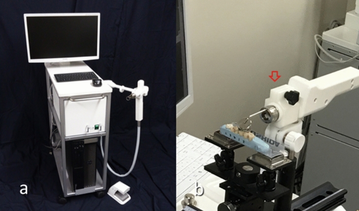
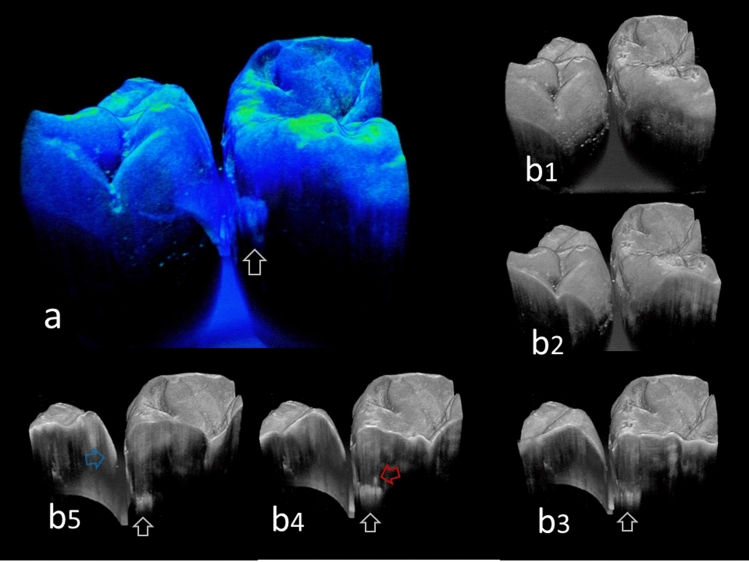
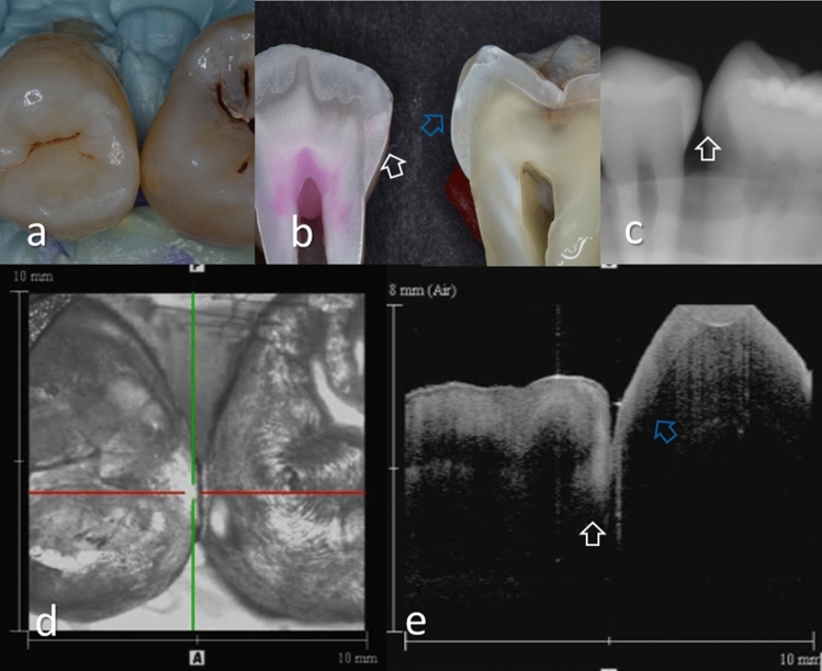
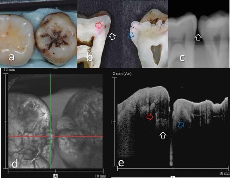
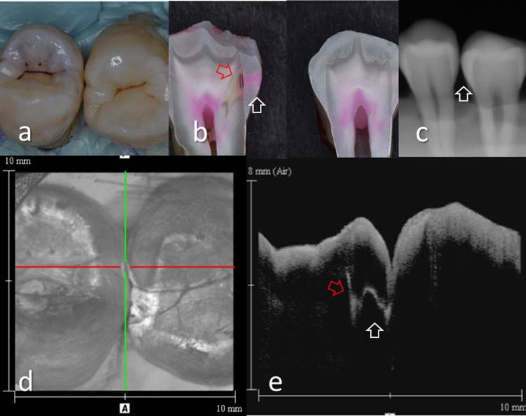
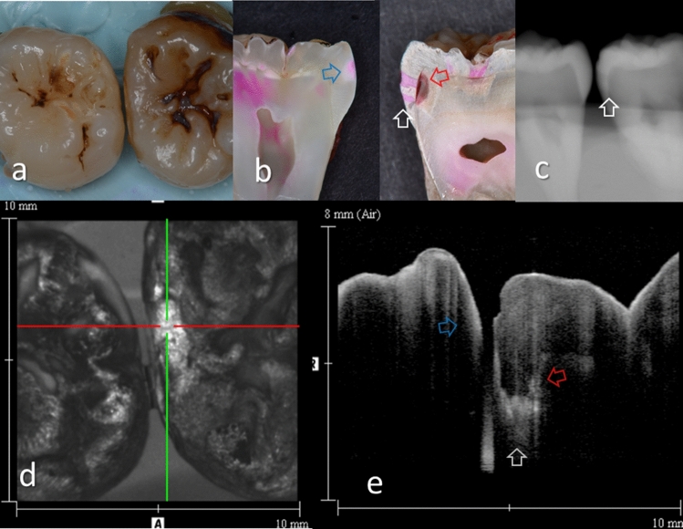
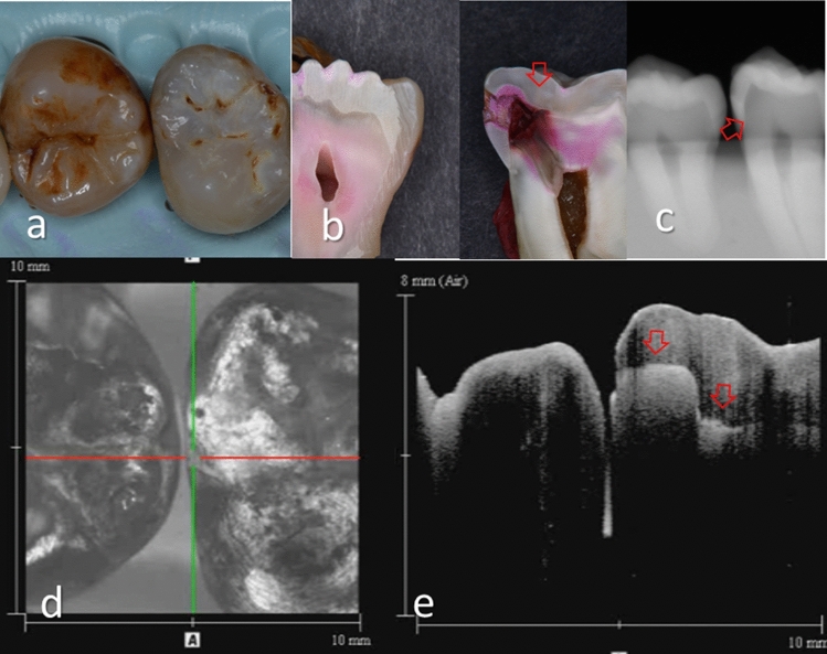
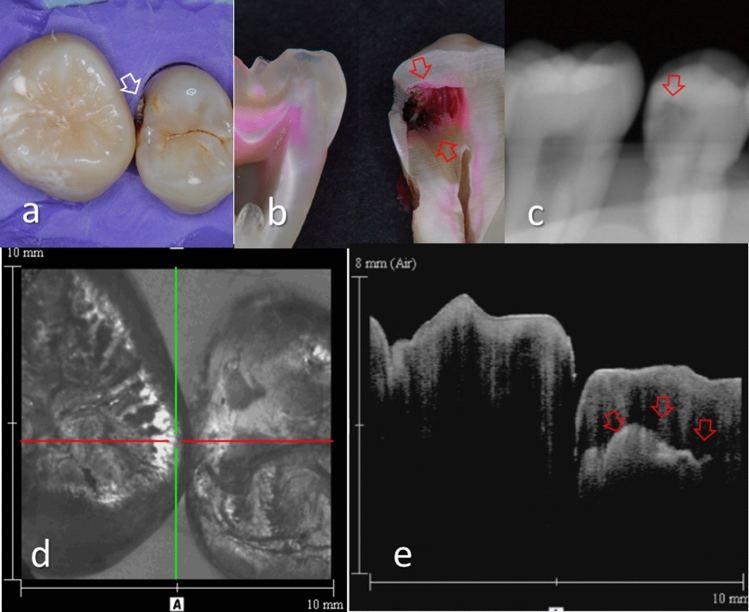
Optical coherence tomography (OCT) can create cross-sectional images of tooth without X-ray exposure. This study aimed to investigate the diagnostic accuracy of 3D imaging of OCT for proximal caries in posterior teeth. Thirty-six human molar teeth with 51 proximal surfaces visibly 6 intact, 16 slightly demineralized, and 29 distinct carious changes were mounted to take digital radiographs and 3D OCT images. The sensitivity, specificity and area under the receiver operating characteristic curve (AUC) for the diagnosis of enamel caries and dentin caries were calculated to quantify the diagnostic ability of 3D OCT in comparison with digital radiography. Diagnostic accuracy was evaluated by the agreement with histology using weighted Kappa. OCT showed significantly higher sensitivity, AUC and Kappa values than radiography. OCT can be a safer option for the diagnosis of proximal caries in posterior teeth that can be applied to the patients without X-ray exposure.
光学相干断层扫描(OCT)可以在不进行 X 射线照射的情况下创建牙齿的横截面图像。本研究旨在探讨 OCT 三维成像对后牙邻面龋的诊断准确性。将 36 个人类磨牙的 51 个近表面分别标记为 6 个完整、16 个轻度脱矿和 29 个明显龋坏,分别进行数字化射线照相和 OCT 三维成像。计算 OCT 对釉质龋和牙本质龋的诊断灵敏度、特异度和受试者工作特征曲线(ROC)下面积(AUC),以量化与数字化射线照相相比,OCT 三维成像的诊断能力。通过与组织学的一致性评估诊断准确性,使用加权 Kappa。OCT 的灵敏度、AUC 和 Kappa 值明显高于射线照相。OCT 可为不进行 X 射线照射的患者提供一种更安全的后牙邻面龋诊断选择。