NIHR Biomedical Research Centre for Ophthalmology, Moorfields Eye Hospital NHS Foundation Trust and UCL Institute of Ophthalmology, London, United Kingdom.
Google Health, London, United Kingdom.
Ophthalmology. 2021 May;128(5):693-705. doi: 10.1016/j.ophtha.2020.09.025. Epub 2020 Sep 24.
To apply a deep learning algorithm for automated, objective, and comprehensive quantification of OCT scans to a large real-world dataset of eyes with neovascular age-related macular degeneration (AMD) and make the raw segmentation output data openly available for further research.
Retrospective analysis of OCT images from the Moorfields Eye Hospital AMD Database.
A total of 2473 first-treated eyes and 493 second-treated eyes that commenced therapy for neovascular AMD between June 2012 and June 2017.
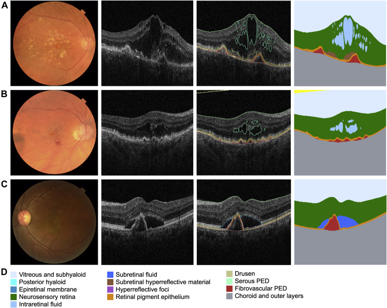
A deep learning algorithm was used to segment all baseline OCT scans. Volumes were calculated for segmented features such as neurosensory retina (NSR), drusen, intraretinal fluid (IRF), subretinal fluid (SRF), subretinal hyperreflective material (SHRM), retinal pigment epithelium (RPE), hyperreflective foci (HRF), fibrovascular pigment epithelium detachment (fvPED), and serous PED (sPED). Analyses included comparisons between first- and second-treated eyes by visual acuity (VA) and race/ethnicity and correlations between volumes.
Volumes of segmented features (mm) and central subfield thickness (CST) (μm).
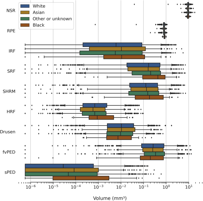
In first-treated eyes, the majority had both IRF and SRF (54.7%). First-treated eyes had greater volumes for all segmented tissues, with the exception of drusen, which was greater in second-treated eyes. In first-treated eyes, older age was associated with lower volumes for RPE, SRF, NSR, and sPED; in second-treated eyes, older age was associated with lower volumes of NSR, RPE, sPED, fvPED, and SRF. Eyes from Black individuals had higher SRF, RPE, and serous PED volumes compared with other ethnic groups. Greater volumes of the majority of features were associated with worse VA.
We report the results of large-scale automated quantification of a novel range of baseline features in neovascular AMD. Major differences between first- and second-treated eyes, with increasing age, and between ethnicities are highlighted. In the coming years, enhanced, automated OCT segmentation may assist personalization of real-world care and the detection of novel structure-function correlations. These data will be made publicly available for replication and future investigation by the AMD research community.
应用深度学习算法对来自真实世界的大量新生血管性年龄相关性黄斑变性(AMD)眼的 OCT 扫描进行自动、客观和全面的定量分析,并公开原始分割输出数据以用于进一步的研究。
回顾性分析 Moorfields 眼科医院 AMD 数据库中的 OCT 图像。
共有 2473 只首次治疗眼和 493 只第二次治疗眼,这些眼于 2012 年 6 月至 2017 年 6 月期间开始接受新生血管性 AMD 的治疗。
使用深度学习算法对所有基线 OCT 扫描进行分割。计算分割后的特征体积,如神经感觉视网膜(NSR)、玻璃膜疣、视网膜内液(IRF)、视网膜下液(SRF)、视网膜下高反射物质(SHRM)、视网膜色素上皮(RPE)、高反射灶(HRF)、纤维血管性色素上皮脱离(fvPED)和浆液性 PED(sPED)。分析包括按视力(VA)和种族/民族比较首次治疗眼和第二次治疗眼,并对体积进行相关性分析。
分割特征的体积(mm)和中央凹下区厚度(CST)(μm)。
在首次治疗眼中,大多数眼同时存在 IRF 和 SRF(54.7%)。与第二次治疗眼相比,首次治疗眼的所有分割组织的体积都更大,除了玻璃膜疣的体积更大。在首次治疗眼中,年龄越大,RPE、SRF、NSR 和 sPED 的体积越小;在第二次治疗眼中,年龄越大,NSR、RPE、sPED、fvPED 和 SRF 的体积越小。与其他种族相比,黑人的 SRF、RPE 和浆液性 PED 体积更高。大多数特征的体积越大,VA 越差。
我们报告了对新生血管性 AMD 的一系列新的基线特征进行大规模自动定量分析的结果。首次治疗眼和第二次治疗眼之间、年龄增长以及种族之间的主要差异被强调。在未来几年,增强的、自动化的 OCT 分割可能有助于个性化真实世界的护理,并检测新的结构-功能相关性。这些数据将公开提供给 AMD 研究界进行复制和进一步研究。