Zhai Xuetong, Santosa Hendrik, Huppert Theodore J
University of Pittsburgh, Department of Bioengineering, Pittsburgh, Pennsylvania, United States.
University of Pittsburgh, Department of Radiology, Pittsburgh, Pennsylvania, United States.
Neurophotonics. 2020 Jul;7(3):035008. doi: 10.1117/1.NPh.7.3.035008. Epub 2020 Sep 23.
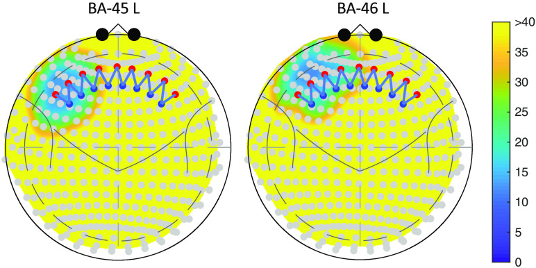
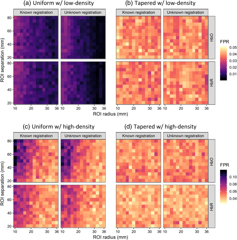
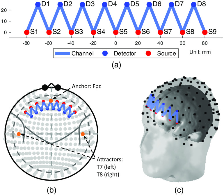
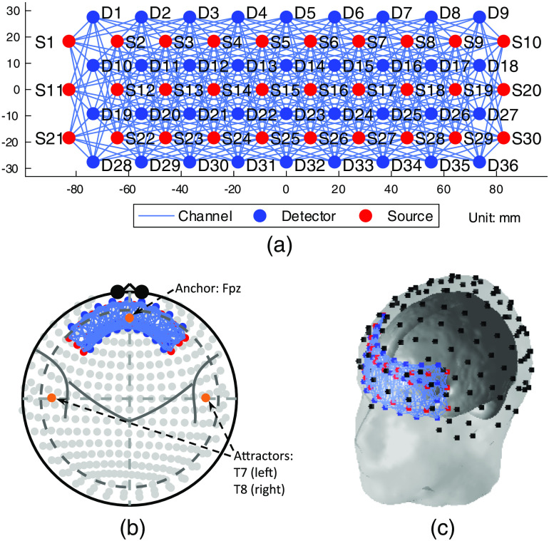
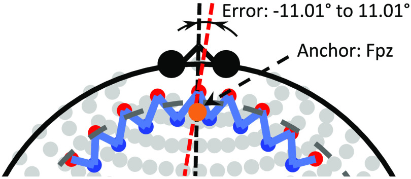
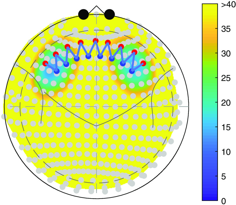
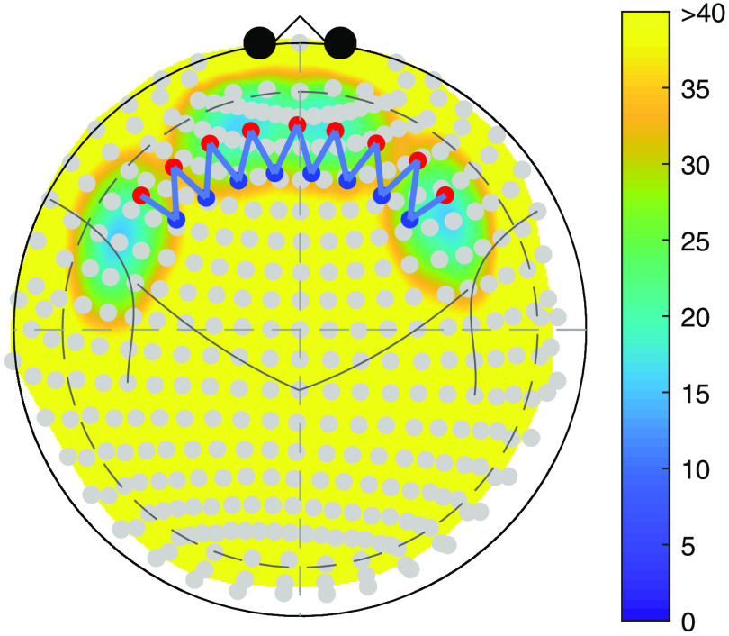
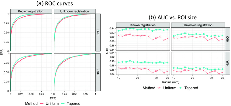
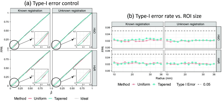
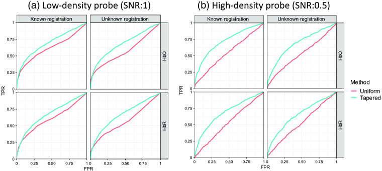
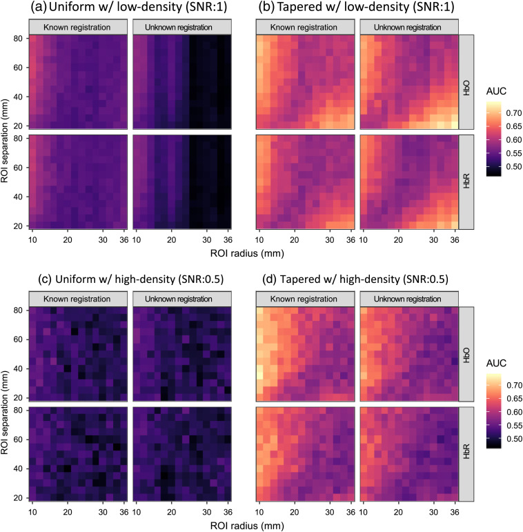
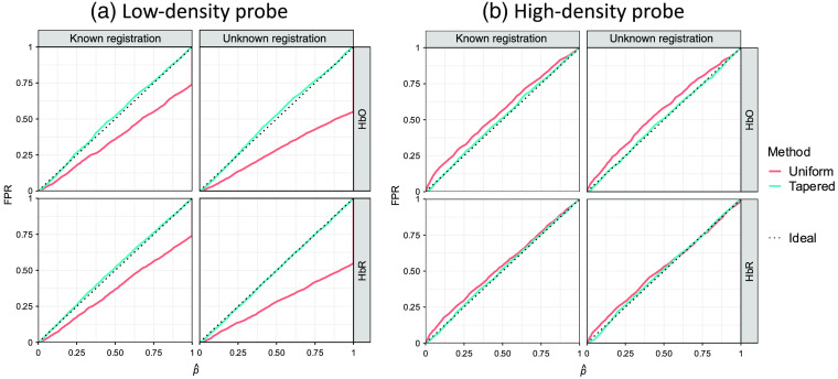
Functional near-infrared spectroscopy (fNIRS) uses surface-placed light sources and detectors to record underlying changes in the brain due to fluctuations in hemoglobin levels and oxygenation. Since these measurements are recorded from the surface of the scalp, the mapping from underlying regions-of-interest (ROIs) in the brain space to the fNIRS channel space measurements depends on the registration of the sensors, the anatomy of the head/brain, and the sensitivity of these diffuse measurements through the tissue. However, small displacements in the probe position can change the distribution of recorded brain activity across the fNIRS measurements. We propose an approach using either individual or atlas-based brain-space anatomical information to define ROI-based statistical hypotheses to test the null involvement of specific regions, which allows us to test the analogous ROI across subjects while adjusting for fNIRS probe placement and sensitivity differences due to head size variations without a localizer task. We use the optical forward model to project the underlying brain-space ROI into a tapered contrast vector, which defines the relative weighting of the fNIRS channels contributing to the ROI and allows us to test the null hypothesis of no brain activity in this region during a functional task. We demonstrate this method through simulation and compare the sensitivity-specificity of this approach to other conventional methods. We examine the performance of this method in the scenario where head size and probe registration are both an accurately known parameters and where this is subject to unknown experimental errors. This method is compared with the performance of the conventional method using 364 different simulation parameter combinations. The proposed method is always recommended in ROI-based analysis, since it significantly improves the analysis performance without a localizer task, wherever the fNIRS probe registration is known or unknown.
功能近红外光谱技术(fNIRS)使用置于头皮表面的光源和探测器,来记录由于血红蛋白水平和氧合作用波动而引起的大脑潜在变化。由于这些测量是从头皮表面进行记录的,因此从大脑空间中潜在的感兴趣区域(ROI)到fNIRS通道空间测量值的映射,取决于传感器的配准、头部/大脑的解剖结构以及这些通过组织的漫射测量的灵敏度。然而,探头位置的微小位移会改变fNIRS测量中记录的大脑活动分布。我们提出一种方法,利用个体或基于图谱的大脑空间解剖信息来定义基于ROI的统计假设,以检验特定区域的零参与度,这使我们能够在调整因头部大小变化导致的fNIRS探头放置和灵敏度差异的同时,在不同受试者之间测试类似的ROI,而无需定位任务。我们使用光学正向模型将潜在的大脑空间ROI投影到一个锥形对比向量中,该向量定义了对ROI有贡献的fNIRS通道的相对权重,并使我们能够在功能任务期间检验该区域无大脑活动的零假设。我们通过模拟演示了这种方法,并将该方法的灵敏度 - 特异性与其他传统方法进行了比较。我们在头部大小和探头配准均为准确已知参数以及存在未知实验误差的情况下,研究了该方法的性能。使用364种不同的模拟参数组合,将该方法与传统方法的性能进行了比较。在基于ROI的分析中,总是推荐使用所提出的方法,因为无论fNIRS探头配准是已知还是未知,该方法都能在无需定位任务的情况下显著提高分析性能。