Department of Biological Chemistry and Molecular Pharmacology, Harvard Medical School, Boston, MA, United States of America.
Astbury Centre for Structural Molecular Biology, School of Molecular & Cellular Biology, Faculty of Biological Sciences, University of Leeds, Leeds, United Kingdom.
PLoS Pathog. 2020 Sep 30;16(9):e1008920. doi: 10.1371/journal.ppat.1008920. eCollection 2020 Sep.
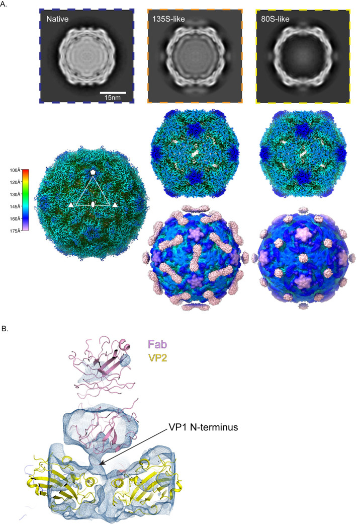
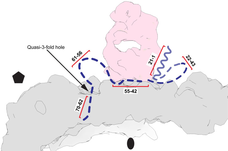
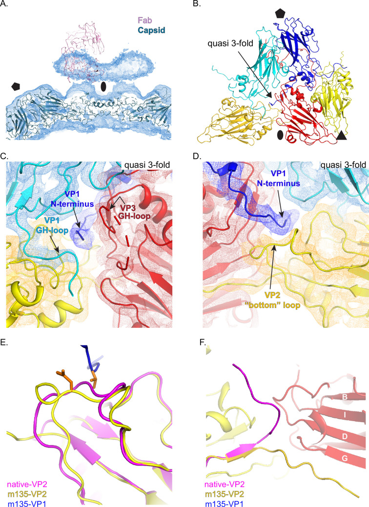
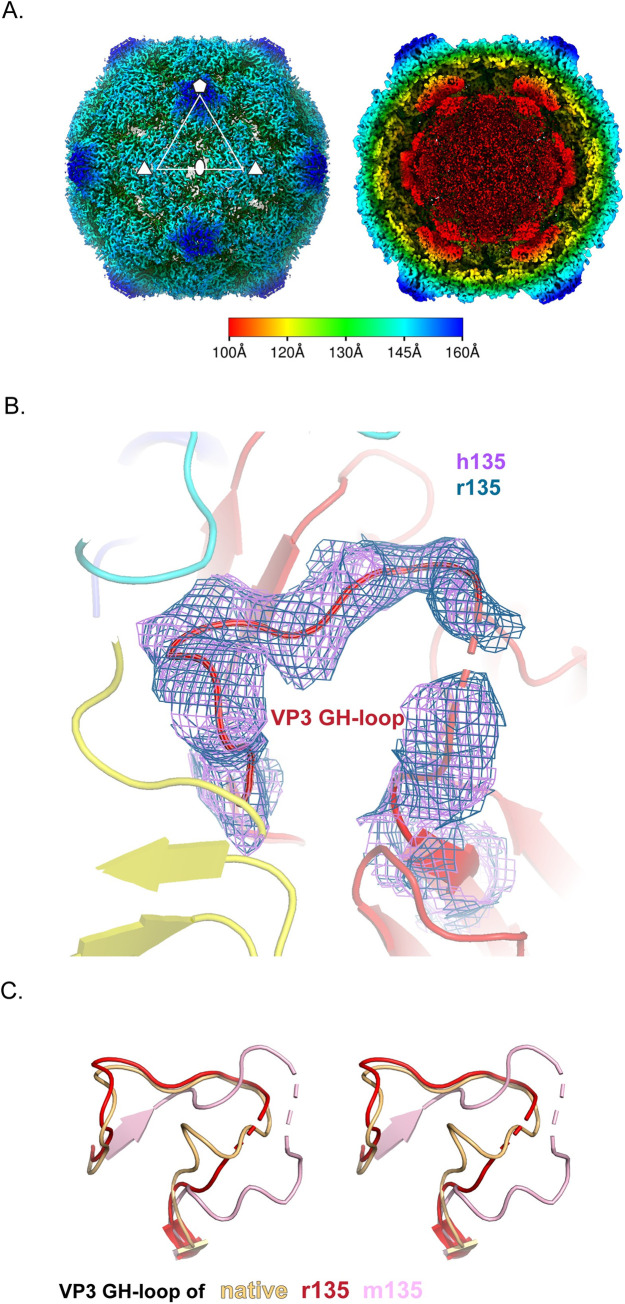
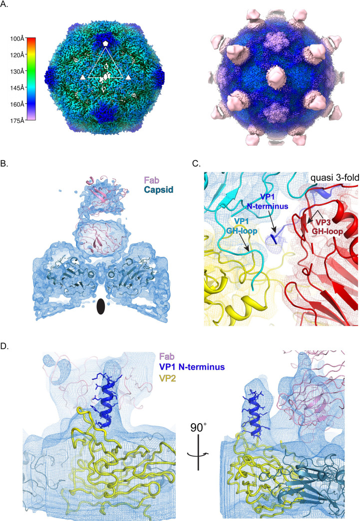
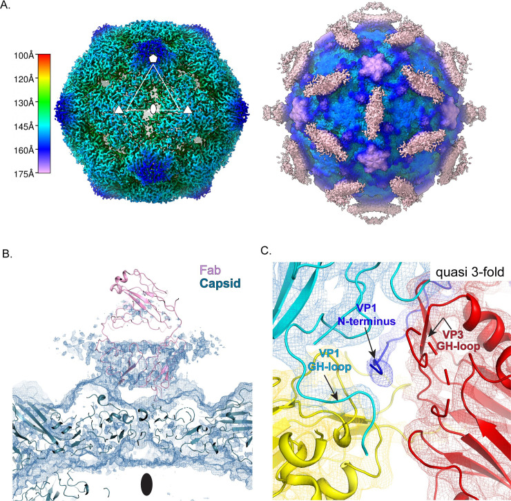
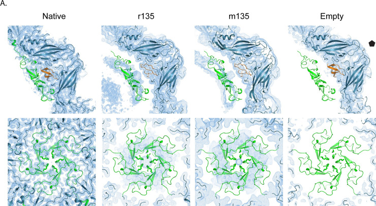
The virions of enteroviruses such as poliovirus undergo a global conformational change after binding to the cellular receptor, characterized by a 4% expansion, and by the opening of holes at the two and quasi-three-fold symmetry axes of the capsid. The resultant particle is called a 135S particle or A-particle and is thought to be on the pathway to a productive infection. Previously published studies have concluded that the membrane-interactive peptides, namely VP4 and the N-terminus of VP1, are irreversibly externalized in the 135S particle. However, using established protocols to produce the 135S particle, and single particle cryo-electron microscopy methods, we have identified at least two unique states that we call the early and late 135S particle. Surprisingly, only in the "late" 135S particles have detectable levels of the VP1 N-terminus been trapped outside the capsid. Moreover, we observe a distinct density inside the capsid that can be accounted for by VP4 that remains associated with the genome. Taken together our results conclusively demonstrate that the 135S particle is not a unique conformation, but rather a family of conformations that could exist simultaneously.
肠道病毒(如脊髓灰质炎病毒)的病毒粒子在与细胞受体结合后会经历全构象变化,其特征是 4%的扩张,并在衣壳的二倍和准三倍对称轴处打开孔。由此产生的颗粒称为 135S 颗粒或 A 颗粒,被认为是通向有效感染的途径。先前的研究已经得出结论,膜相互作用肽,即 VP4 和 VP1 的 N 端,在 135S 颗粒中不可逆地向外延伸。然而,使用既定的方案来产生 135S 颗粒,并采用单颗粒冷冻电子显微镜方法,我们鉴定了至少两种独特的状态,我们称之为早期和晚期 135S 颗粒。令人惊讶的是,只有在“晚期”135S 颗粒中,才能检测到衣壳外可探测水平的 VP1 N 端。此外,我们观察到衣壳内有一个明显的密度,可以由与基因组相关联的 VP4 来解释。总之,我们的结果明确表明,135S 颗粒不是一种独特的构象,而是可能同时存在的构象家族。