• 文献检索
  • 文档翻译
  • 深度研究
  • 学术资讯
  • Suppr Zotero 插件Zotero 插件
  • 邀请有礼
  • 套餐&价格
  • 历史记录
应用&插件
Suppr Zotero 插件Zotero 插件浏览器插件Mac 客户端Windows 客户端微信小程序
定价
高级版会员购买积分包购买API积分包
服务
文献检索文档翻译深度研究API 文档MCP 服务
关于我们
关于 Suppr公司介绍联系我们用户协议隐私条款
关注我们

Suppr 超能文献

核心技术专利:CN118964589B侵权必究
粤ICP备2023148730 号-1Suppr @ 2026

文献检索

告别复杂PubMed语法,用中文像聊天一样搜索,搜遍4000万医学文献。AI智能推荐,让科研检索更轻松。

立即免费搜索

文件翻译

保留排版,准确专业,支持PDF/Word/PPT等文件格式,支持 12+语言互译。

免费翻译文档

深度研究

AI帮你快速写综述,25分钟生成高质量综述,智能提取关键信息,辅助科研写作。

立即免费体验

FalseColor-Python:用于基于荧光的无载玻片数字病理学的快速强度均衡和数字染色包。

FalseColor-Python: A rapid intensity-leveling and digital-staining package for fluorescence-based slide-free digital pathology.

机构信息

Department of Mechanical Engineering, University of Washington, Seattle, Washington, United States of America.

Department of Pathology, University of Washington, Seattle, Washington, United States of America.

出版信息

PLoS One. 2020 Oct 1;15(10):e0233198. doi: 10.1371/journal.pone.0233198. eCollection 2020.

DOI:10.1371/journal.pone.0233198
PMID:33001995
原文链接:https://pmc.ncbi.nlm.nih.gov/articles/PMC7529223/
Abstract

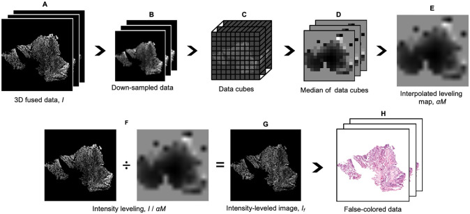
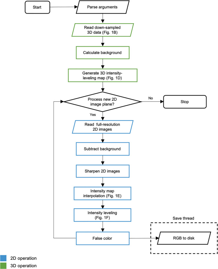
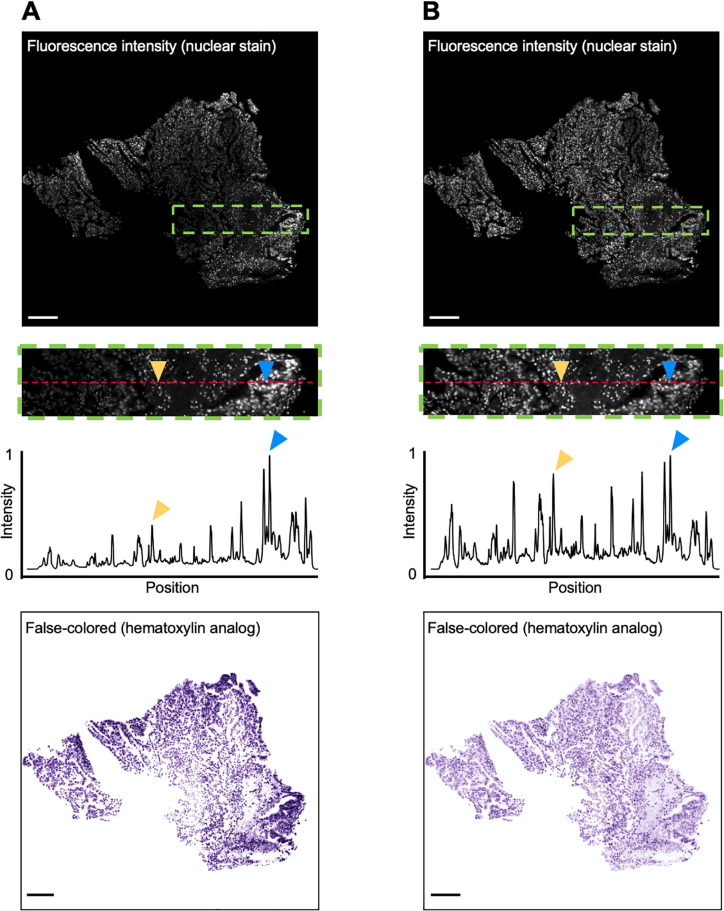
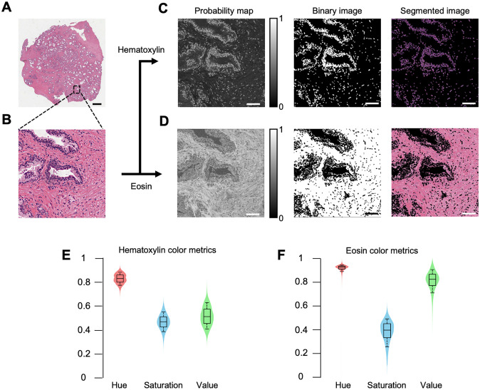
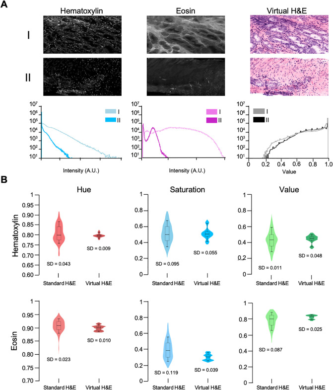
Slide-free digital pathology techniques, including nondestructive 3D microscopy, are gaining interest as alternatives to traditional slide-based histology. In order to facilitate clinical adoption of these fluorescence-based techniques, software methods have been developed to convert grayscale fluorescence images into color images that mimic the appearance of standard absorptive chromogens such as hematoxylin and eosin (H&E). However, these false-coloring algorithms often require manual and iterative adjustment of parameters, with results that can be inconsistent in the presence of intensity nonuniformities within an image and/or between specimens (intra- and inter-specimen variability). Here, we present an open-source (Python-based) rapid intensity-leveling and digital-staining package that is specifically designed to render two-channel fluorescence images (i.e. a fluorescent analog of H&E) to the traditional H&E color space for 2D and 3D microscopy datasets. However, this method can be easily tailored for other false-coloring needs. Our package offers (1) automated and uniform false coloring in spite of uneven staining within a large thick specimen, (2) consistent color-space representations that are robust to variations in staining and imaging conditions between different specimens, and (3) GPU-accelerated data processing to allow these methods to scale to large datasets. We demonstrate this platform by generating H&E-like images from cleared tissues that are fluorescently imaged in 3D with open-top light-sheet (OTLS) microscopy, and quantitatively characterizing the results in comparison to traditional slide-based H&E histology.

摘要

无滑动物镜技术,包括无损 3D 显微镜,正作为传统载玻片组织学的替代方法受到关注。为了促进这些基于荧光的技术在临床中的应用,已经开发了软件方法将灰度荧光图像转换为模仿标准吸收性染料(如苏木精和伊红(H&E))外观的彩色图像。然而,这些伪彩色算法通常需要手动和迭代调整参数,并且在图像内和/或标本之间存在强度不均匀性的情况下,结果可能不一致(标本内和标本间变异性)。在这里,我们提出了一个开源(基于 Python)的快速强度均衡和数字染色包,专门用于将双通道荧光图像(即 H&E 的荧光类似物)渲染到传统的 H&E 彩色空间,用于 2D 和 3D 显微镜数据集。然而,这种方法可以很容易地适应其他伪彩色需求。我们的软件包提供了(1)即使在大厚标本内存在不均匀染色的情况下也能实现自动和均匀的伪彩色,(2)对不同标本之间染色和成像条件变化具有稳健的一致颜色空间表示,以及(3)GPU 加速的数据处理,以使这些方法能够扩展到大型数据集。我们通过从用开放式顶光片(OTLS)显微镜荧光成像的 3D 透明组织生成 H&E 样图像来展示该平台,并与传统的基于载玻片的 H&E 组织学相比对结果进行定量表征。

https://cdn.ncbi.nlm.nih.gov/pmc/blobs/23f3/7529223/75dccf99bcb3/pone.0233198.g006.jpg
https://cdn.ncbi.nlm.nih.gov/pmc/blobs/23f3/7529223/3de64b2b1059/pone.0233198.g001.jpg
https://cdn.ncbi.nlm.nih.gov/pmc/blobs/23f3/7529223/25a27c1c571b/pone.0233198.g002.jpg
https://cdn.ncbi.nlm.nih.gov/pmc/blobs/23f3/7529223/fdfe8bc3c469/pone.0233198.g003.jpg
https://cdn.ncbi.nlm.nih.gov/pmc/blobs/23f3/7529223/5c3e55de2197/pone.0233198.g004.jpg
https://cdn.ncbi.nlm.nih.gov/pmc/blobs/23f3/7529223/0953c3fbc784/pone.0233198.g005.jpg
https://cdn.ncbi.nlm.nih.gov/pmc/blobs/23f3/7529223/75dccf99bcb3/pone.0233198.g006.jpg

相似文献

1
FalseColor-Python: A rapid intensity-leveling and digital-staining package for fluorescence-based slide-free digital pathology.FalseColor-Python:用于基于荧光的无载玻片数字病理学的快速强度均衡和数字染色包。
PLoS One. 2020 Oct 1;15(10):e0233198. doi: 10.1371/journal.pone.0233198. eCollection 2020.
2
Open-Top Light-Sheet Microscopy Image Atlas of Prostate Core Needle Biopsies.前列腺核心针活检的开放式光片显微镜图像图谱。
Arch Pathol Lab Med. 2019 Sep;143(9):1069-1075. doi: 10.5858/arpa.2018-0466-OA. Epub 2019 Mar 20.
3
Axially swept open-top light-sheet microscopy for densely labeled clinical specimens.轴向扫掠开顶式光片显微镜用于高密度标记的临床标本。
Opt Lett. 2024 Jul 1;49(13):3794-3797. doi: 10.1364/OL.521591.
4
A Guide to Perform 3D Histology of Biological Tissues with Fluorescence Microscopy.《荧光显微镜下生物组织 3D 组织学操作指南》
Int J Mol Sci. 2023 Apr 4;24(7):6747. doi: 10.3390/ijms24076747.
5
An end-to-end workflow for nondestructive 3D pathology.用于无损 3D 病理学的端到端工作流程。
Nat Protoc. 2024 Apr;19(4):1122-1148. doi: 10.1038/s41596-023-00934-4. Epub 2024 Jan 23.
6
Nondestructive 3D Pathology Image Atlas of Barrett Esophagus With Open-Top Light-Sheet Microscopy.采用开顶式光片显微镜的 Barrett 食管无损 3D 病理学图像图谱。
Arch Pathol Lab Med. 2023 Oct 1;147(10):1164-1171. doi: 10.5858/arpa.2022-0133-OA.
7
Comparing Accuracy of Helicobacter pylori Identification Using Traditional Hematoxylin and Eosin-Stained Glass Slides With Digital Whole Slide Imaging.比较传统苏木精-伊红染色玻片与数字全玻片成像在幽门螺杆菌鉴定中的准确性。
Lab Invest. 2024 Jan;104(1):100262. doi: 10.1016/j.labinv.2023.100262. Epub 2023 Oct 13.
8
Applications and challenges of digital pathology and whole slide imaging.数字病理学与全切片成像的应用及挑战
Biotech Histochem. 2015 Jul;90(5):341-7. doi: 10.3109/10520295.2015.1044566. Epub 2015 May 15.
9
A Pilot Validation Study Comparing Fluorescence-Imitating Brightfield Imaging, A Slide-Free Imaging Method, With Standard Formalin-Fixed, Paraffin-Embedded Hematoxylin-Eosin-Stained Tissue Section Histology for Primary Surgical Pathology Diagnosis.荧光模拟明场成像与免载玻片成像方法与标准福尔马林固定石蜡包埋苏木精-伊红染色组织切片组织病理学用于原发性外科病理诊断的初步验证性研究
Arch Pathol Lab Med. 2024 Mar 1;148(3):345-352. doi: 10.5858/arpa.2022-0432-OA.
10
Conventional histological and cytological staining with simultaneous immunohistochemistry enabled by invisible chromogens.常规的组织学和细胞学染色与不可见显色剂的同时免疫组织化学相结合。
Lab Invest. 2022 May;102(5):545-553. doi: 10.1038/s41374-021-00714-2. Epub 2021 Dec 28.

引用本文的文献

1
Deep-learning triage of 3D pathology datasets for comprehensive and efficient pathologist assessments.用于全面高效的病理学家评估的3D病理数据集的深度学习分类
bioRxiv. 2025 Jul 22:2025.07.20.665804. doi: 10.1101/2025.07.20.665804.
2
Vaginal biomechanical function in premenopausal and postmenopausal women with and without pelvic organ prolapse.有和没有盆腔器官脱垂的绝经前和绝经后女性的阴道生物力学功能。
Sci Rep. 2025 Jul 25;15(1):27039. doi: 10.1038/s41598-025-11094-8.
3
Investigating hepatic fibrosis heterogeneity by three-dimensional imaging in metabolic dysfunction-associated steatotic liver disease.

本文引用的文献

1
Stop Explaining Black Box Machine Learning Models for High Stakes Decisions and Use Interpretable Models Instead.停止为高风险决策解释黑箱机器学习模型,转而使用可解释模型。
Nat Mach Intell. 2019 May;1(5):206-215. doi: 10.1038/s42256-019-0048-x. Epub 2019 May 13.
2
BigStitcher: reconstructing high-resolution image datasets of cleared and expanded samples.BigStitcher:重建已清除和扩展样本的高分辨率图像数据集。
Nat Methods. 2019 Sep;16(9):870-874. doi: 10.1038/s41592-019-0501-0. Epub 2019 Aug 5.
3
Multi-immersion open-top light-sheet microscope for high-throughput imaging of cleared tissues.
通过三维成像研究代谢功能障碍相关脂肪性肝病中的肝纤维化异质性
Sci Rep. 2025 Jul 1;15(1):20736. doi: 10.1038/s41598-025-98680-y.
4
Exploring Multiplex Immunohistochemistry (mIHC) Techniques and Histopathology Image Analysis: Current Practice and Potential for Clinical Incorporation.探索多重免疫组织化学(mIHC)技术与组织病理学图像分析:当前实践与临床应用潜力
Cancer Med. 2025 Jan;14(1):e70523. doi: 10.1002/cam4.70523.
5
ML-driven segmentation of microvascular features during histological examination of tissue-engineered vascular grafts.基于机器学习的组织工程血管移植物组织学检查中微血管特征分割
Front Bioeng Biotechnol. 2024 Jun 26;12:1411680. doi: 10.3389/fbioe.2024.1411680. eCollection 2024.
6
descSPIM: an affordable and easy-to-build light-sheet microscope optimized for tissue clearing techniques.descSPIM:一种经济实惠且易于构建的光片显微镜,针对组织透明化技术进行了优化。
Nat Commun. 2024 Jun 12;15(1):4941. doi: 10.1038/s41467-024-49131-1.
7
Analysis of 3D pathology samples using weakly supervised AI.使用弱监督 AI 分析 3D 病理样本。
Cell. 2024 May 9;187(10):2502-2520.e17. doi: 10.1016/j.cell.2024.03.035.
8
Direct three-dimensional segmentation of prostate glands with nnU-Net.使用nnU-Net对前列腺进行直接三维分割。
J Biomed Opt. 2024 Mar;29(3):036001. doi: 10.1117/1.JBO.29.3.036001. Epub 2024 Mar 1.
9
An end-to-end workflow for nondestructive 3D pathology.用于无损 3D 病理学的端到端工作流程。
Nat Protoc. 2024 Apr;19(4):1122-1148. doi: 10.1038/s41596-023-00934-4. Epub 2024 Jan 23.
10
Engineering the future of 3D pathology.工程化 3D 病理学的未来。
J Pathol Clin Res. 2024 Jan;10(1):e347. doi: 10.1002/cjp2.347. Epub 2023 Nov 2.
多浸开放式光片显微镜,用于高通量成像透明化组织。
Nat Commun. 2019 Jul 4;10(1):2781. doi: 10.1038/s41467-019-10534-0.
4
Virtual histological staining of unlabelled tissue-autofluorescence images via deep learning.基于深度学习的无标记组织自发荧光图像的虚拟组织学染色。
Nat Biomed Eng. 2019 Jun;3(6):466-477. doi: 10.1038/s41551-019-0362-y. Epub 2019 Mar 4.
5
Microscopy with ultraviolet surface excitation for rapid slide-free histology.紫外表面激发显微镜,快速免载玻片组织学。
Nat Biomed Eng. 2017 Dec;1(12):957-966. doi: 10.1038/s41551-017-0165-y. Epub 2017 Dec 4.
6
Comparing histologic evaluation of prostate tissue using nonlinear microscopy and paraffin H&E: a pilot study.使用非线性显微镜和石蜡 H&E 对前列腺组织进行组织学评估的比较:一项初步研究。
Mod Pathol. 2019 Jul;32(8):1158-1167. doi: 10.1038/s41379-019-0250-8. Epub 2019 Mar 26.
7
Open-Top Light-Sheet Microscopy Image Atlas of Prostate Core Needle Biopsies.前列腺核心针活检的开放式光片显微镜图像图谱。
Arch Pathol Lab Med. 2019 Sep;143(9):1069-1075. doi: 10.5858/arpa.2018-0466-OA. Epub 2019 Mar 20.
8
Digital staining through the application of deep neural networks to multi-modal multi-photon microscopy.通过将深度神经网络应用于多模态多光子显微镜进行数字染色。
Biomed Opt Express. 2019 Feb 19;10(3):1339-1350. doi: 10.1364/BOE.10.001339. eCollection 2019 Mar 1.
9
Tissue Clearing and Light Sheet Microscopy: Imaging the Unsectioned Adult Zebra Finch Brain at Cellular Resolution.组织透明化与光片显微镜技术:以细胞分辨率对未切片的成年斑胸草雀大脑进行成像
Front Neuroanat. 2019 Feb 14;13:13. doi: 10.3389/fnana.2019.00013. eCollection 2019.
10
Microscopy with ultraviolet surface excitation for wide-area pathology of breast surgical margins.紫外表面激发显微镜用于乳房外科切缘的大面积病理学检查。
J Biomed Opt. 2019 Feb;24(2):1-11. doi: 10.1117/1.JBO.24.2.026501.