• 文献检索
  • 文档翻译
  • 深度研究
  • 学术资讯
  • Suppr Zotero 插件Zotero 插件
  • 邀请有礼
  • 套餐&价格
  • 历史记录
应用&插件
Suppr Zotero 插件Zotero 插件浏览器插件Mac 客户端Windows 客户端微信小程序
定价
高级版会员购买积分包购买API积分包
服务
文献检索文档翻译深度研究API 文档MCP 服务
关于我们
关于 Suppr公司介绍联系我们用户协议隐私条款
关注我们

Suppr 超能文献

核心技术专利:CN118964589B侵权必究
粤ICP备2023148730 号-1Suppr @ 2026

文献检索

告别复杂PubMed语法,用中文像聊天一样搜索,搜遍4000万医学文献。AI智能推荐,让科研检索更轻松。

立即免费搜索

文件翻译

保留排版,准确专业,支持PDF/Word/PPT等文件格式,支持 12+语言互译。

免费翻译文档

深度研究

AI帮你快速写综述,25分钟生成高质量综述,智能提取关键信息,辅助科研写作。

立即免费体验

紫外表面激发显微镜,快速免载玻片组织学。

Microscopy with ultraviolet surface excitation for rapid slide-free histology.

机构信息

Department of Pathology and Laboratory Medicine, University of California Davis Medical Center, Sacramento, CA, USA.

Department of Biochemistry and Molecular Medicine, University of California Davis Medical Center, Sacramento, CA, USA.

出版信息

Nat Biomed Eng. 2017 Dec;1(12):957-966. doi: 10.1038/s41551-017-0165-y. Epub 2017 Dec 4.

DOI:10.1038/s41551-017-0165-y
PMID:31015706
原文链接:https://pmc.ncbi.nlm.nih.gov/articles/PMC6223324/
Abstract

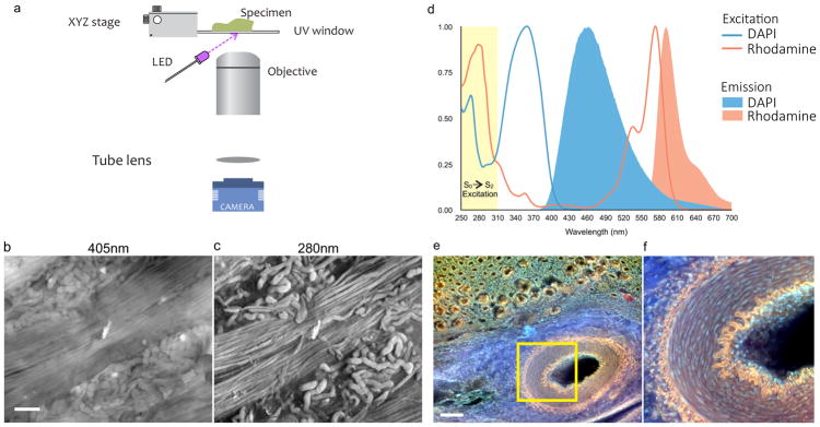
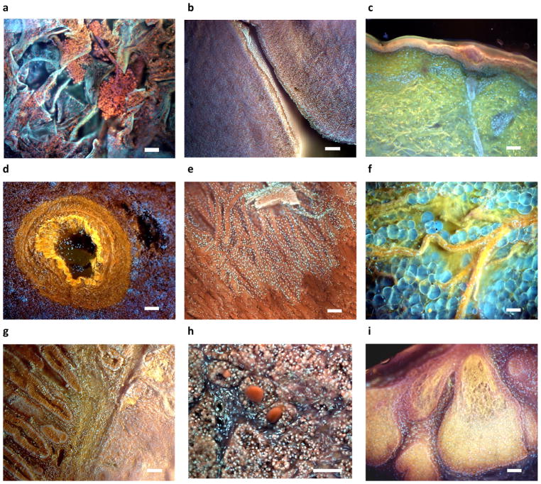
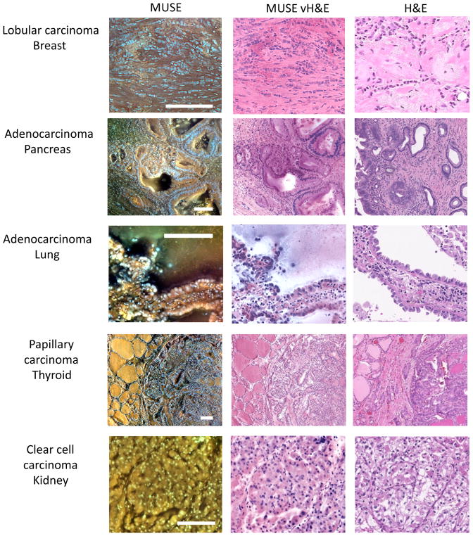
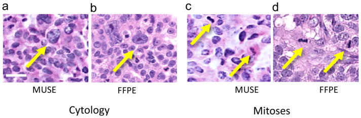
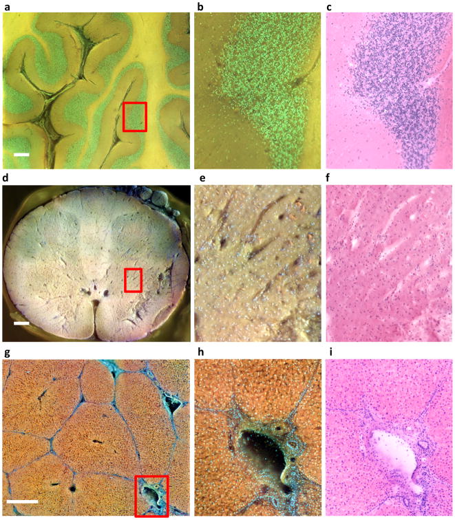
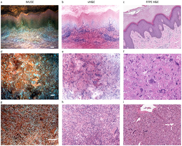
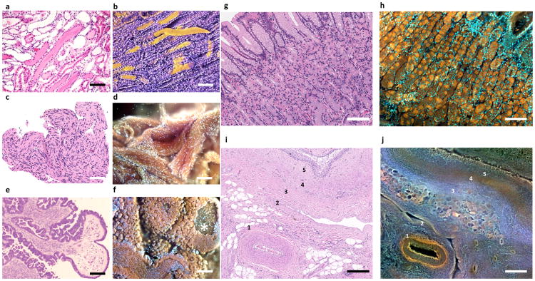
Histological examination of tissues is central to the diagnosis and management of neoplasms and many other diseases and is a foundational technique for preclinical and basic research. However, commonly used bright-field microscopy requires prior preparation of micrometre-thick tissue sections mounted on glass slides-a process that can require hours or days, contributes to cost and delays access to critical information. Here, we introduce a simple, non-destructive slide-free technique that, within minutes, provides high-resolution diagnostic histological images resembling those obtained from conventional haematoxylin and eosin histology. The approach, which we named microscopy with ultraviolet surface excitation (MUSE), can also generate shape and colour-contrast information. MUSE relies on ~280 nm ultraviolet light to restrict the excitation of conventional fluorescent stains to tissue surfaces and it has no significant effects on downstream molecular assays (including fluorescence in situ hybridization and RNA sequencing). MUSE promises to improve the speed and efficiency of patient care in both state-of-the-art and low-resource settings and to provide opportunities for rapid histology in research.

摘要

组织的组织学检查是肿瘤和许多其他疾病的诊断和治疗的核心,也是临床前和基础研究的基础技术。然而,常用的明场显微镜需要预先制备微米厚的组织切片,这些切片安装在载玻片上,这个过程可能需要数小时或数天,不仅增加了成本,还延迟了获取关键信息的时间。在这里,我们介绍了一种简单、非破坏性的免载玻片技术,它可以在几分钟内提供类似于传统苏木精和伊红染色的高分辨率诊断组织学图像。我们将这种方法命名为紫外表面激发显微镜(MUSE),它还可以生成形状和颜色对比信息。MUSE 依赖于~280nm 的紫外光将传统荧光染料的激发限制在组织表面,对下游分子检测(包括荧光原位杂交和 RNA 测序)没有显著影响。MUSE 有望提高在先进和资源匮乏环境中患者护理的速度和效率,并为研究中的快速组织学提供机会。

https://cdn.ncbi.nlm.nih.gov/pmc/blobs/27c0/6223324/2bc7bbf56a0c/nihms916826f7.jpg
https://cdn.ncbi.nlm.nih.gov/pmc/blobs/27c0/6223324/c401b8113325/nihms916826f1.jpg
https://cdn.ncbi.nlm.nih.gov/pmc/blobs/27c0/6223324/48d6144fccd7/nihms916826f2.jpg
https://cdn.ncbi.nlm.nih.gov/pmc/blobs/27c0/6223324/98373fc19dfc/nihms916826f3.jpg
https://cdn.ncbi.nlm.nih.gov/pmc/blobs/27c0/6223324/e5e09f7f80c6/nihms916826f4.jpg
https://cdn.ncbi.nlm.nih.gov/pmc/blobs/27c0/6223324/81bf382aa219/nihms916826f5.jpg
https://cdn.ncbi.nlm.nih.gov/pmc/blobs/27c0/6223324/5356886b87b8/nihms916826f6.jpg
https://cdn.ncbi.nlm.nih.gov/pmc/blobs/27c0/6223324/2bc7bbf56a0c/nihms916826f7.jpg

相似文献

1
Microscopy with ultraviolet surface excitation for rapid slide-free histology.紫外表面激发显微镜,快速免载玻片组织学。
Nat Biomed Eng. 2017 Dec;1(12):957-966. doi: 10.1038/s41551-017-0165-y. Epub 2017 Dec 4.
2
Rapid histopathological imaging of skin and breast cancer surgical specimens using immersion microscopy with ultraviolet surface excitation.利用紫外表面激发浸液显微镜对皮肤和乳腺癌手术标本进行快速组织病理学成像。
Sci Rep. 2018 Mar 14;8(1):4476. doi: 10.1038/s41598-018-22264-2.
3
Microscopy with ultraviolet surface excitation for wide-area pathology of breast surgical margins.紫外表面激发显微镜用于乳房外科切缘的大面积病理学检查。
J Biomed Opt. 2019 Feb;24(2):1-11. doi: 10.1117/1.JBO.24.2.026501.
4
Microscopy with ultraviolet surface excitation (MUSE): A novel approach to real-time inexpensive slide-free dermatopathology.紫外线表面激发显微镜检查(MUSE):一种用于实时、低成本无玻片皮肤病理学检查的新方法。
J Cutan Pathol. 2018 Jul;45(7):498-503. doi: 10.1111/cup.13255. Epub 2018 May 8.
5
Deep-learning-assisted microscopy with ultraviolet surface excitation for rapid slide-free histological imaging.用于快速无玻片组织学成像的紫外表面激发深度学习辅助显微镜技术。
Biomed Opt Express. 2021 Sep 1;12(9):5920-5938. doi: 10.1364/BOE.433597.
6
Fast and slide-free imaging.快速且无滑动成像。
Nat Biomed Eng. 2017 Dec;1(12):926-928. doi: 10.1038/s41551-017-0172-z.
7
Critical comparison of 31 commercially available digital slide systems in pathology.31种市售病理学数字切片系统的关键比较
Int J Surg Pathol. 2006 Oct;14(4):285-305. doi: 10.1177/1066896906292274.
8
Adaptation of microscopy with ultraviolet surface excitation for enhancing STEM and undergraduate education.采用紫外表面激发显微镜技术增强 STEM 并促进本科教育
J Biomed Opt. 2018 Jul;23(12):1-8. doi: 10.1117/1.JBO.23.12.121603.
9
Simultaneous deep ultraviolet transmission and scattering microscopy for virtual histology.用于虚拟组织学的同时深紫外透射和散射显微镜。
Opt Lett. 2024 May 15;49(10):2729-2732. doi: 10.1364/OL.514077.
10
Autofocus evaluation for brightfield microscopy pathology.明场显微镜病理学的自动对焦评估。
J Biomed Opt. 2012 Mar;17(3):036008. doi: 10.1117/1.JBO.17.3.036008.

引用本文的文献

1
Label-free surface sectioning deep ultraviolet tissue imaging in multimodalities.多模态无标记表面切片深紫外组织成像
Biomed Opt Express. 2025 Jun 16;16(7):2756-2766. doi: 10.1364/BOE.565070. eCollection 2025 Jul 1.
2
Rapid full-color serial sectioning tomography with speckle illumination and ultraviolet excitation.采用散斑照明和紫外线激发的快速全彩连续切片断层扫描
Npj Imaging. 2024 Sep 30;2(1):37. doi: 10.1038/s44303-024-00040-4.
3
Fast and label-free 3D virtual H&E histology via active phase modulation-assisted dynamic full-field OCT.

本文引用的文献

1
Rapid intraoperative histology of unprocessed surgical specimens via fibre-laser-based stimulated Raman scattering microscopy.通过基于光纤激光的受激拉曼散射显微镜对未处理手术标本进行快速术中组织学检查。
Nat Biomed Eng. 2017;1. doi: 10.1038/s41551-016-0027. Epub 2017 Feb 6.
2
Current State of the Regulatory Trajectory for Whole Slide Imaging Devices in the USA.美国全切片成像设备监管轨迹的现状
J Pathol Inform. 2017 May 15;8:23. doi: 10.4103/jpi.jpi_11_17. eCollection 2017.
3
Real-time, High-resolution, In Vivo Characterization of Superficial Skin With Microscopy Using Ultraviolet Surface Excitation (MUSE).
通过主动相位调制辅助动态全场光学相干断层扫描实现快速且无标记的三维虚拟苏木精-伊红组织学成像
Npj Imaging. 2025 Apr 8;3(1):12. doi: 10.1038/s44303-025-00068-0.
4
H&E Style Translation Using CycleGAN for Deep Ultraviolet-Excitation Fluorescence Images of Pancreatic Endoscopic Ultrasound-Fine Needle Aspiration/Biopsy Toward Slide-Free Rapid Pathology.使用循环生成对抗网络(CycleGAN)对胰腺内镜超声细针穿刺/活检的深紫外激发荧光图像进行苏木精-伊红(H&E)风格转换以实现无玻片快速病理学诊断
Acta Histochem Cytochem. 2025 Apr 26;58(2):59-67. doi: 10.1267/ahc.25-00007. Epub 2025 Apr 9.
5
Emerging Trends in Point-of-Care Technology Development for Oncology in Low- and Middle-Income Countries.低收入和中等收入国家肿瘤学即时护理技术发展的新趋势
JCO Glob Oncol. 2025 Jun;11:e2500142. doi: 10.1200/GO-25-00142. Epub 2025 Jun 18.
6
Toward real-time margin assessment in breast-conserving surgery with hyperspectral imaging.利用高光谱成像技术实现保乳手术中的实时切缘评估
Sci Rep. 2025 Mar 20;15(1):9556. doi: 10.1038/s41598-025-94526-9.
7
Visualization of Gastric Adenocarcinoma Lymph Node Metastases by Microscopy with Ultraviolet Surface Excitation.通过紫外线表面激发显微镜观察胃腺癌淋巴结转移情况
Sovrem Tekhnologii Med. 2024;16(6):25-32. doi: 10.17691/stm2024.16.6.03. Epub 2024 Dec 27.
8
Quantifying UV-induced photodamage for longitudinal live-cell imaging applications of deep-UV microscopy.用于深紫外显微镜纵向活细胞成像应用的紫外线诱导光损伤定量分析。
Biomed Opt Express. 2024 Dec 19;16(1):208-221. doi: 10.1364/BOE.544778. eCollection 2025 Jan 1.
9
Optical-resolution parallel ultraviolet photoacoustic microscopy for slide-free histology.用于无玻片组织学的光学分辨率并行紫外光声显微镜。
Sci Adv. 2024 Dec 13;10(50):eado0518. doi: 10.1126/sciadv.ado0518. Epub 2024 Dec 11.
10
Label- and slide-free tissue histology using 3D epi-mode quantitative phase imaging and virtual hematoxylin and eosin staining.使用3D落射模式定量相成像和虚拟苏木精与伊红染色的无标记和无玻片组织组织学
Optica. 2023 Dec 20;10(12):1605-1618. doi: 10.1364/optica.502859. Epub 2023 Dec 1.
使用紫外线表面激发显微镜(MUSE)对浅表皮肤进行实时、高分辨率的体内表征。
J Drugs Dermatol. 2016 Nov 1;15(11):1344-1346.
4
Measure transcript integrity using RNA-seq data.使用RNA测序数据测量转录本完整性。
BMC Bioinformatics. 2016 Feb 3;17:58. doi: 10.1186/s12859-016-0922-z.
5
New Colors for Histology: Optimized Bivariate Color Maps Increase Perceptual Contrast in Histological Images.组织学的新色彩:优化的双变量色图增强组织学图像中的感知对比度
PLoS One. 2015 Dec 30;10(12):e0145572. doi: 10.1371/journal.pone.0145572. eCollection 2015.
6
Swept confocally-aligned planar excitation (SCAPE) microscopy for high speed volumetric imaging of behaving organisms.用于行为生物体高速体积成像的扫描共焦对齐平面激发(SCAPE)显微镜。
Nat Photonics. 2015 Feb;9(2):113-119. doi: 10.1038/nphoton.2014.323.
7
Variation in pre-PCR processing of FFPE samples leads to discrepancies in BRAF and EGFR mutation detection: a diagnostic RING trial.福尔马林固定石蜡包埋(FFPE)样本的PCR前处理差异导致BRAF和EGFR突变检测结果出现差异:一项诊断性RING试验
J Clin Pathol. 2015 Feb;68(2):111-8. doi: 10.1136/jclinpath-2014-202644. Epub 2014 Nov 27.
8
Confocal fluorescence microscopy for rapid evaluation of invasive tumor cellularity of inflammatory breast carcinoma core needle biopsies.共聚焦荧光显微镜用于快速评估炎性乳腺癌粗针活检的浸润性肿瘤细胞密度。
Breast Cancer Res Treat. 2015 Jan;149(1):303-10. doi: 10.1007/s10549-014-3182-5. Epub 2014 Nov 23.
9
Assessment of breast pathologies using nonlinear microscopy.使用非线性显微镜评估乳腺病变
Proc Natl Acad Sci U S A. 2014 Oct 28;111(43):15304-9. doi: 10.1073/pnas.1416955111. Epub 2014 Oct 13.
10
Pathology of inherited manganese transporter deficiency.遗传性锰转运体缺乏症的病理学。
Ann Neurol. 2014 Apr;75(4):608-12. doi: 10.1002/ana.24131. Epub 2014 Apr 14.