From the Departments of Pathology (Drs Reder, True, and Liu and Ms McCarty) and Mechanical Engineering (Drs Glaser and Liu and Mr Chen), University of Washington, Seattle.
Arch Pathol Lab Med. 2019 Sep;143(9):1069-1075. doi: 10.5858/arpa.2018-0466-OA. Epub 2019 Mar 20.
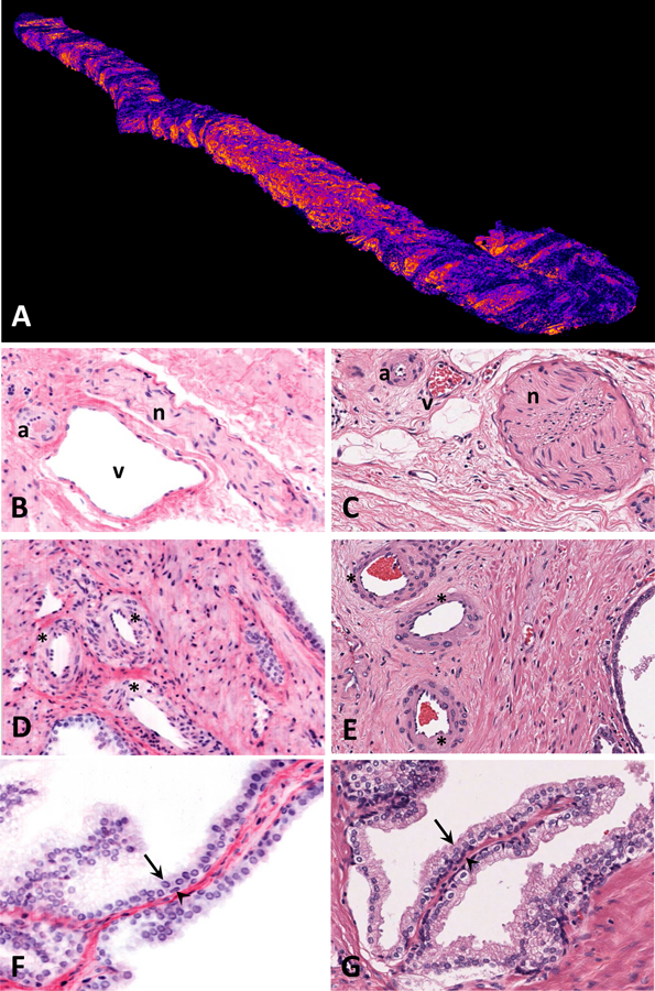
CONTEXT.—: Ex vivo microscopy encompasses a range of techniques to examine fresh or fixed tissue with microscopic resolution, eliminating the need to embed the tissue in paraffin or produce a glass slide. One such technique is light-sheet microscopy, which enables rapid 3D imaging. Our pathology-engineering collaboration has resulted in an open-top light-sheet (OTLS) microscope that is specifically tailored to the needs of pathology practice.
OBJECTIVE.—: To present an image atlas of OTLS images of prostate core needle biopsies.
DESIGN.—: Core needle biopsies (N = 9) were obtained from fresh radical prostatectomy specimens. Each biopsy was fixed in formalin, dehydrated in ethanol, stained with TO-PRO3 and eosin, optically cleared, and imaged using OTLS microscopy. The biopsies were then processed, paraffin embedded, and sectioned. Hematoxylin-eosin and immunohistochemical staining for cytokeratin 5 and cytokeratin 8 was performed.
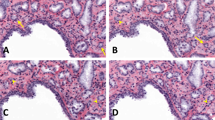
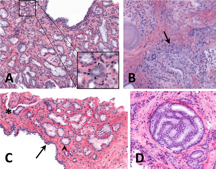
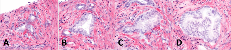
RESULTS.—: Benign and neoplastic histologic structures showed high fidelity between OTLS and traditional light microscopy. OTLS microscopy had no discernible effect on hematoxylin-eosin or immunohistochemical staining in this pilot study. The 3D histology information obtained using OTLS microscopy enabled new structural insights, including the observation of cribriform and well-formed gland morphologies within the same contiguous glandular structures, as well as the continuity of poorly formed glands with well-formed glands.
CONCLUSIONS.—: Three-dimensional OTLS microscopy images have a similar appearance to traditional hematoxylin-eosin histology images, with the added benefit of useful 3D structural information. Further studies are needed to continue to document the OTLS appearance of a wide range of tissues and to better understand 3D histologic structures.
离体显微镜技术涵盖了一系列用于检查新鲜或固定组织的微观分辨率的技术,无需将组织嵌入石蜡中或制作玻璃载玻片。其中一种技术是光片显微镜,它可以实现快速的 3D 成像。我们的病理学-工程合作产生了一种专门针对病理学实践需求的开放式光片(OTLS)显微镜。
展示前列腺核心针活检的 OTLS 图像图谱。
从新鲜的根治性前列腺切除术标本中获得核心针活检(N=9)。每个活检都用福尔马林固定,用乙醇脱水,用 TO-PRO3 和曙红染色,光学透明化,并使用 OTLS 显微镜进行成像。然后对活检进行处理、石蜡包埋和切片。进行苏木精-伊红和细胞角蛋白 5 和细胞角蛋白 8 的免疫组织化学染色。
良性和肿瘤组织学结构在 OTLS 和传统光学显微镜之间具有高度的保真度。在这项初步研究中,OTLS 显微镜对苏木精-伊红或免疫组织化学染色没有明显影响。使用 OTLS 显微镜获得的 3D 组织学信息提供了新的结构见解,包括在同一连续腺结构内观察到筛状和形态良好的腺体形态,以及形态不规则的腺体与形态良好的腺体的连续性。
三维 OTLS 显微镜图像与传统的苏木精-伊红组织学图像具有相似的外观,并且具有有用的 3D 结构信息的额外优势。需要进一步的研究来继续记录 OTLS 对广泛组织的外观,并更好地了解 3D 组织学结构。