Zong Min, Zhao Hua, Li Qiang, Li Yanbing, Zhang Jianjun
Department of Cardiology, Beijing Chaoyang Hospital, Capital Medical University, Beijing 100043, P.R. China.
Exp Ther Med. 2020 Nov;20(5):117. doi: 10.3892/etm.2020.9245. Epub 2020 Sep 18.
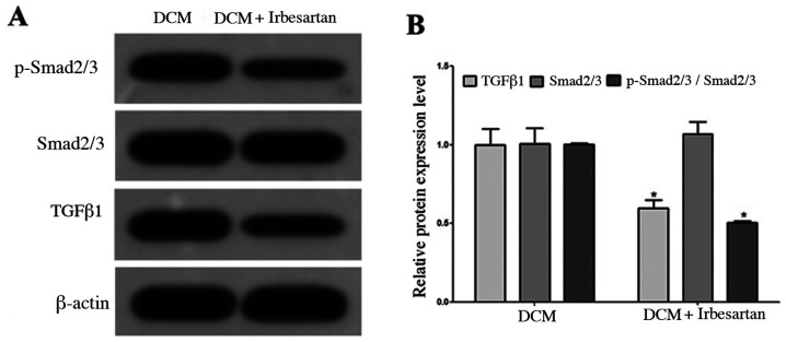
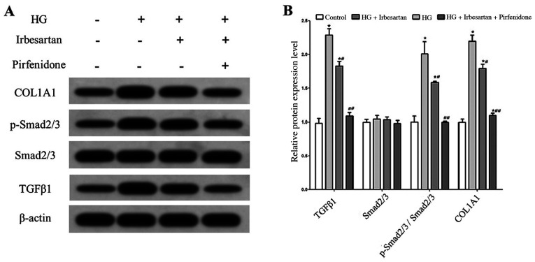
Myocardial fibrosis (MF) is an important pathological change in diabetic cardiomyopathy. The aim of the present study was to investigate whether irbesartan serves a role in improving MF in a diabetic rat model. Fasting blood glucose (FBG), total cholesterol (TC), triglyceride (TG), high-density lipoprotein cholesterol (HDL-C) and low-density lipoprotein cholesterol (LDL-C) levels were measured in rats using biochemical methods. Heart weight index (HWI), left ventricular weight index (LVWI), left ventricular systolic pressure (LVSP) and left ventricular end-diastolic pressure (LVEDP) were also measured, whilst type I collagen and hydroxyproline content in myocardial tissue was quantified. Western blotting was used to measure the expression of transforming growth factor β1 (TGFβ1), phosphorylated (p)-Smad2/3 and collagen type I α 1 chain (COL1A1) inmyocardial tissues or rat cardiac fibroblast (RCF) cells. Cell proliferation was measured using EdU staining. Procollagen type III N-terminal peptide (PIIINP) content, FBG, TC, TG and LDL-C levels were found to be significantly higher, whilst HDL-C levels were found to be significantly lower in rats in the diabetic group. Those in the diabetic group also exhibited significantly elevated HWI, LVWI, LVEDP, myocardial tissue type I collagen content and hydroxyproline content values, but significantly reduced LVSP. Changes in the aforementioned indicators were reversed after treatment with irbesartan, where the protein expression levels of TGFβ1 and p-Smad2/3 in myocardial tissue were also significantly reduced. In RCF cells, irbesartan significantly reversed high glucose-induced upregulation of TGFβ1 expression, Smad2/3 phosphorylation and COL1A1 expression, as well as reducing cell proliferation and rat type I PICP and PIIINP levels. Application of pirfenidone produced additive effects on reducing the expression levels of the proteins aforementioned when combined with irbesartan. Therefore, the present results demonstrated that irbesartan reduced the activity of the TGFβ1/Smad2/3 pathway and ameliorated diabetic MF by downregulating the expression of TGFβ1.
心肌纤维化(MF)是糖尿病性心肌病的一种重要病理变化。本研究的目的是探讨厄贝沙坦在改善糖尿病大鼠模型的MF中是否起作用。采用生化方法测定大鼠空腹血糖(FBG)、总胆固醇(TC)、甘油三酯(TG)、高密度脂蛋白胆固醇(HDL-C)和低密度脂蛋白胆固醇(LDL-C)水平。还测量了心脏重量指数(HWI)、左心室重量指数(LVWI)、左心室收缩压(LVSP)和左心室舒张末期压力(LVEDP),同时对心肌组织中的I型胶原蛋白和羟脯氨酸含量进行了定量分析。采用蛋白质印迹法检测心肌组织或大鼠心脏成纤维细胞(RCF)中转化生长因子β1(TGFβ1)、磷酸化(p)-Smad2/3和I型胶原α-1链(COL1A1)的表达。使用EdU染色法检测细胞增殖情况。结果发现,糖尿病组大鼠的III型前胶原N端肽(PIIINP)含量、FBG、TC、TG和LDL-C水平显著升高,而HDL-C水平显著降低。糖尿病组大鼠的HWI、LVWI、LVEDP、心肌组织I型胶原蛋白含量和羟脯氨酸含量值也显著升高,但LVSP显著降低。厄贝沙坦治疗后,上述指标的变化得到逆转,心肌组织中TGFβ1和p-Smad2/3的蛋白表达水平也显著降低。在RCF细胞中,厄贝沙坦显著逆转了高糖诱导的TGFβ1表达上调、Smad2/3磷酸化和COL1A1表达,同时减少了细胞增殖以及大鼠I型前胶原羧基端前肽(PICP)和PIIINP水平。与厄贝沙坦联合应用时,吡非尼酮对降低上述蛋白质的表达水平产生了相加作用。因此,本研究结果表明,厄贝沙坦通过下调TGFβ1的表达降低了TGFβ1/Smad2/3信号通路的活性,并改善了糖尿病性MF。