Szczerba Agata, Kuwana Takashi, Paradowska Michelle, Bednarczyk Marek
Department of Animal Biotechnology and Genetics, Faculty of Animal Breeding and Biology, UTP University of Science and Technology, Mazowiecka 28, 85-084 Bydgoszcz, Poland.
Animals (Basel). 2020 Sep 30;10(10):1769. doi: 10.3390/ani10101769.
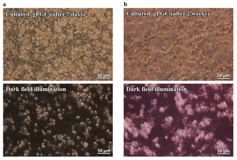
The present study had two aims: (1) To develop a culture system that imitates a normal physiological environment of primordial germ cells (PGCs). There are two types of PGCs in chicken: Circulating blood (cPGCs) and gonadal (gPGCs). The culture condition must support the proliferation of both cPGCs and gPGCs, without affecting their migratory properties and must be deprived of xenobiotic factors, and (2) to propose an easy-to-train, nonlabeling optical technique for the routine identification of live PGCs. To address the first aim, early chicken embryo's feeder cells were examined instead of using feeder cells from mammalian species. The KAv-1 medium at pH 8.0 with the addition of bFGF (basic fibroblast growth factor) was used instead of a conventional culture medium (pH approximately 7.2). Both cPGCs and gPGCs proliferated in vitro and retained their migratory ability after 2 weeks of culture. The cultivated cPGCs and gPGCs colonized the right and/or left gonads of the recipient male and female embryos. To address the second aim, we demonstrated a simple and rapid method to identify live PGCs as bright cells under darkfield illumination. The PGCs rich in lipid droplets in their cytoplasm highly contrasted with the co-cultured feeder layer and other cell populations in the culture.
(1)开发一种模拟原始生殖细胞(PGCs)正常生理环境的培养系统。鸡体内有两种类型的PGCs:循环血液中的PGCs(cPGCs)和性腺中的PGCs(gPGCs)。培养条件必须支持cPGCs和gPGCs的增殖,不影响它们的迁移特性,且必须不含外源性因子;(2)提出一种易于操作、无需标记的光学技术,用于常规鉴定活的PGCs。为实现第一个目的,研究人员检测了早期鸡胚的饲养细胞,而非使用哺乳动物来源的饲养细胞。使用pH值为8.0并添加了碱性成纤维细胞生长因子(bFGF)的KAv-1培养基,而非传统的培养基(pH约为7.2)。cPGCs和gPGCs在体外均能增殖,且在培养2周后仍保留其迁移能力。培养的cPGCs和gPGCs定殖于受体雄性和雌性胚胎的右侧和/或左侧性腺。为实现第二个目的,我们展示了一种简单快速的方法,即在暗视野照明下将活的PGCs鉴定为明亮的细胞。细胞质中富含脂滴的PGCs与共培养的饲养层及培养中的其他细胞群体形成了高度对比。