Mesulam Center for Cognitive Neurology and Alzheimer's Disease, Northwestern University Feinberg School of Medicine, Chicago, IL.
Department of Preventive Medicine, Northwestern University Feinberg School of Medicine, Chicago, IL.
Brain Pathol. 2021 Jan;31(1):189-204. doi: 10.1111/bpa.12902. Epub 2020 Nov 5.
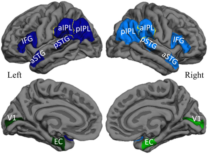
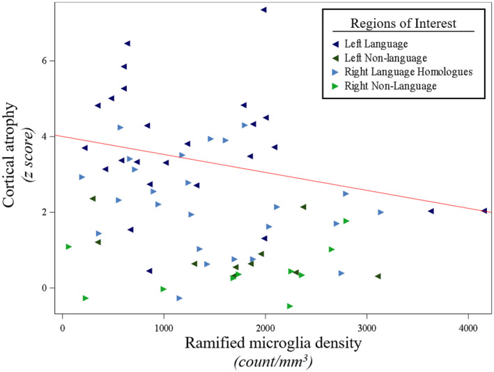
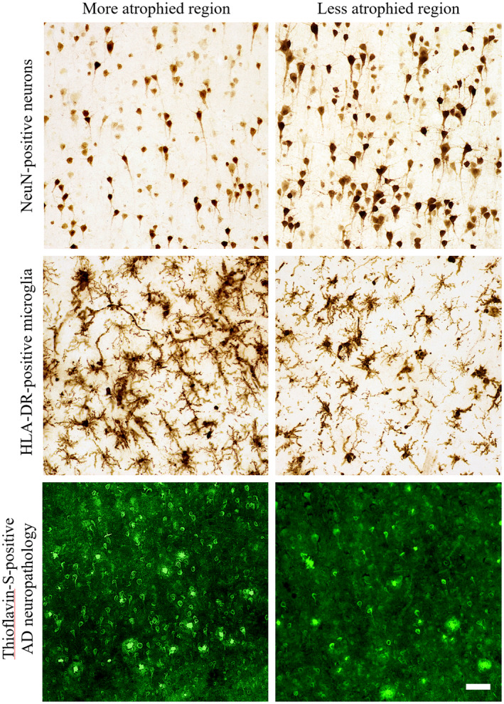
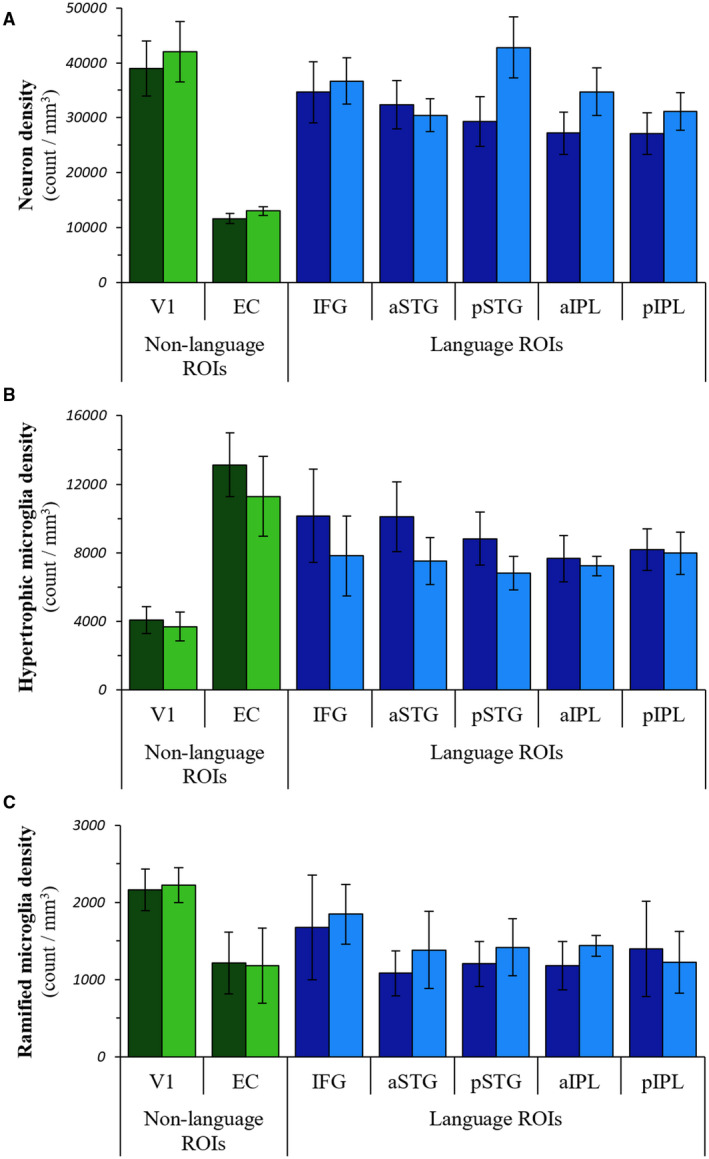
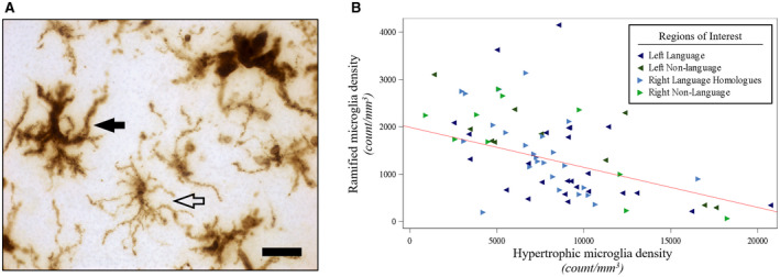
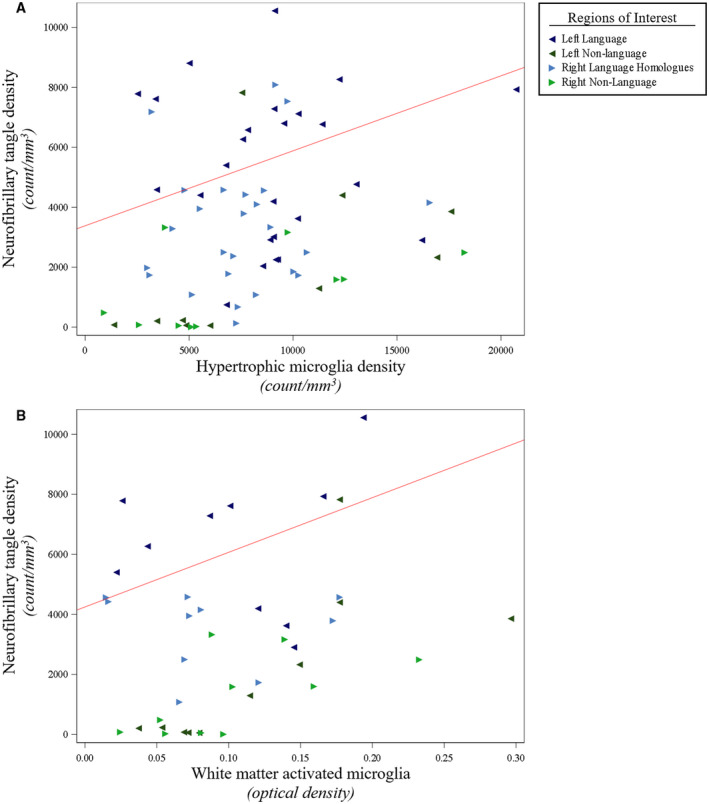
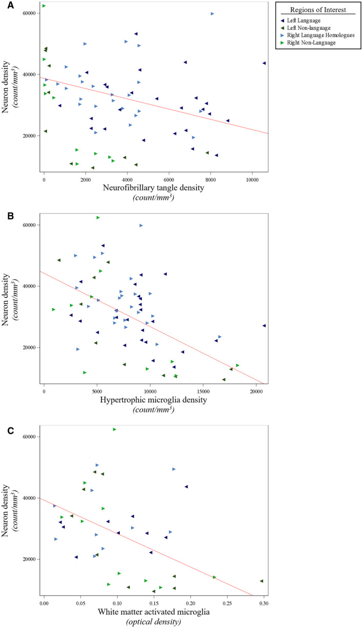
The neurofibrillary tangles (NFT) and amyloid-ß plaques (AP) that comprise Alzheimer's disease (AD) neuropathology are associated with neurodegeneration and microglial activation. Activated microglia exist on a dynamic spectrum of morphologic subtypes that include resting, surveillant microglia capable of converting to activated, hypertrophic microglia closely linked to neuroinflammatory processes and AD neuropathology in amnestic AD. However, quantitative analyses of microglial subtypes and neurons are lacking in non-amnestic clinical AD variants, including primary progressive aphasia (PPA-AD). PPA-AD is a language disorder characterized by cortical atrophy and NFT densities concentrated to the language-dominant hemisphere. Here, a stereologic investigation of five PPA-AD participants determined the densities and distributions of neurons and microglial subtypes to examine how cellular changes relate to AD neuropathology and may contribute to cortical atrophy. Adjacent series of sections were immunostained for neurons (NeuN) and microglia (HLA-DR) from bilateral language and non-language regions where in vivo cortical atrophy and Thioflavin-S-positive APs and NFTs were previously quantified. NeuN-positive neurons and morphologic subtypes of HLA-DR-positive microglia (i.e., resting [ramified] microglia and activated [hypertrophic] microglia) were quantified using unbiased stereology. Relationships between neurons, microglia, AD neuropathology, and cortical atrophy were determined using linear mixed models. NFT densities were positively associated with hypertrophic microglia densities (P < 0.01) and inversely related to neuron densities (P = 0.01). Hypertrophic microglia densities were inversely related to densities of neurons (P < 0.01) and ramified microglia (P < 0.01). Ramified microglia densities were positively associated with neuron densities (P = 0.02) and inversely related to cortical atrophy (P = 0.03). Our findings provide converging evidence of divergent roles for microglial subtypes in patterns of neurodegeneration, which includes hypertrophic microglia likely driving a neuroinflammatory response more sensitive to NFTs than APs in PPA-AD. Moreover, the accumulation of both NFTs and activated hypertrophic microglia in association with low neuron densities suggest they may collectively contribute to focal neurodegeneration characteristic of PPA-AD.
神经原纤维缠结(NFT)和淀粉样-β斑块(AP)组成了阿尔茨海默病(AD)的神经病理学,与神经退行性变和小胶质细胞激活有关。激活的小胶质细胞存在于一个动态的形态亚型谱上,包括静止的、监视的小胶质细胞,它们可以转化为与神经炎症过程和 AD 神经病理学密切相关的活化、肥大的小胶质细胞。然而,在非遗忘型 AD 临床变异型中,包括原发性进行性失语症(PPA-AD),缺乏对小胶质细胞亚型和神经元的定量分析。PPA-AD 是一种以皮质萎缩和 NFT 密度集中在语言优势半球为特征的语言障碍。在这里,对五名 PPA-AD 参与者进行了立体学研究,确定了神经元和小胶质细胞亚型的密度和分布,以研究细胞变化如何与 AD 神经病理学相关,并可能导致皮质萎缩。使用无偏立体学方法,对来自双侧语言和非语言区域的相邻系列切片进行神经元(NeuN)和小胶质细胞(HLA-DR)的免疫染色,这些区域之前已经量化了体内皮质萎缩、硫黄素-S 阳性 AP 和 NFT。使用无偏立体学方法对 NeuN 阳性神经元和 HLA-DR 阳性小胶质细胞的形态亚型(即静止[分支]小胶质细胞和活化[肥大]小胶质细胞)进行定量。使用线性混合模型确定神经元、小胶质细胞、AD 神经病理学和皮质萎缩之间的关系。NFT 密度与肥大小胶质细胞密度呈正相关(P<0.01),与神经元密度呈负相关(P=0.01)。肥大小胶质细胞密度与神经元密度(P<0.01)和分支小胶质细胞密度(P<0.01)呈负相关。分支小胶质细胞密度与神经元密度呈正相关(P=0.02),与皮质萎缩呈负相关(P=0.03)。我们的研究结果提供了小胶质细胞亚型在神经退行性变模式中作用不同的趋同证据,包括肥大小胶质细胞可能比 PPA-AD 中的 AP 更敏感地驱动神经炎症反应。此外,NFT 和活化的肥大小胶质细胞的积累与低神经元密度有关,这表明它们可能共同导致 PPA-AD 特征性的局灶性神经退行性变。