Wang Jiejie, Swanson Raymond A
Department of Neurology, University of California, San Francisco, and San Francisco Veterans Affairs Health Care System, San Francisco, CA, United States.
Front Neurosci. 2020 Sep 3;4:861. doi: 10.3389/fnins.2020.00861. eCollection 2020.
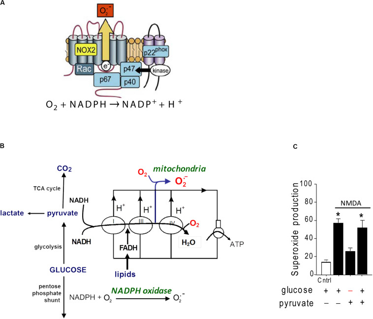
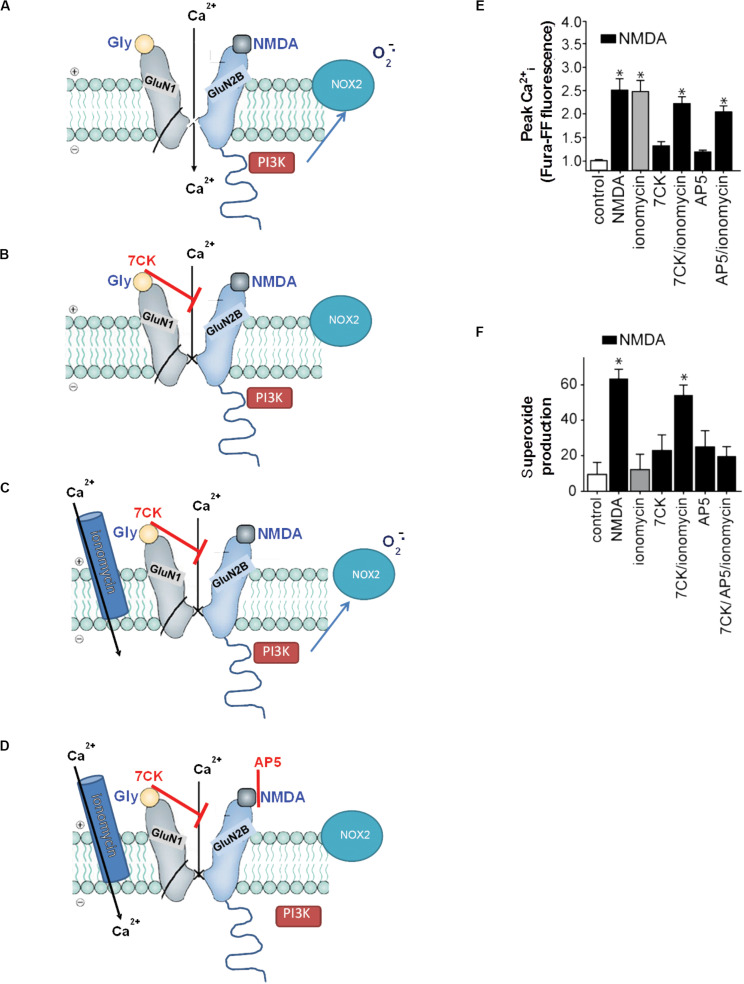
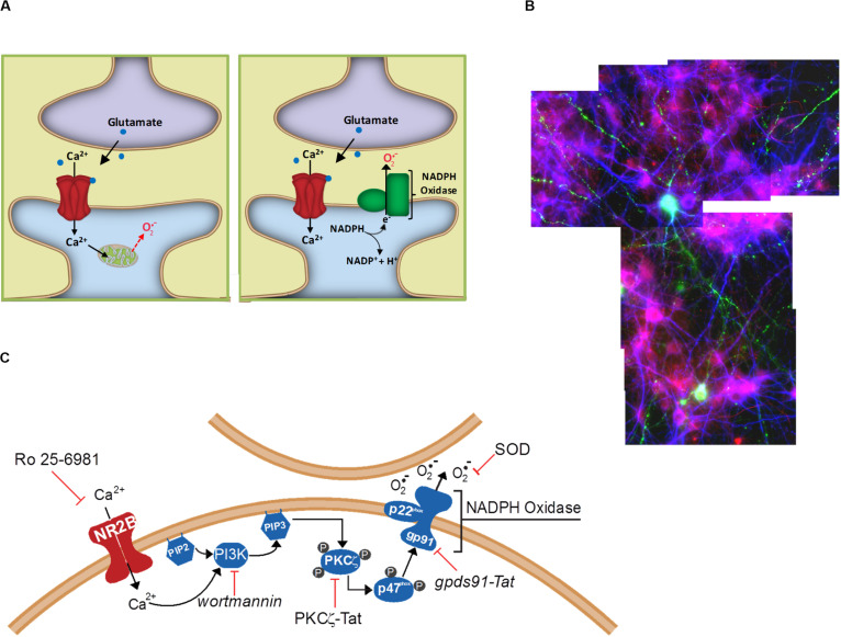
Excitotoxicity is classically attributed to Ca influx through NMDA receptors (NMDAr), leading to production of nitric oxide by neuronal nitric oxide synthase and superoxide by mitochondria, which react to form highly cytotoxic peroxynitrite. More recent observations warrant revision of the classic view and help to explain some otherwise puzzling aspects of excitotoxic cell injury. Studies using pharmacological and genetic approaches show that superoxide produced by NMDAr activation originates primarily from NADPH oxidase rather than from mitochondria. As NADPH oxidase is localized to the plasma membrane, this also provides an explanation for the extracellular release of superoxide and cell-to-cell "spread" of excitotoxic injury observed and . The signaling pathway linking NMDAr to NADPH oxidase involves Ca influx, phosphoinositol-3-kinase, and protein kinase Cζ, and interventions at any of these steps can prevent superoxide production and excitotoxic injury. Ca influx specifically through NMDAr is normally required to induce excitotoxicity, through a mechanism presumed to involve privileged Ca access to local signaling domains. However, experiments using selective blockade of the NMDAr ion channel and artificial reconstitution of Ca by other routes indicate that the special effects of NMDAr activation are attributable instead to concurrent non-ionotropic NMDAr signaling by agonist binding to NMDAr. The non-ionotropic signaling driving NADPH oxidase activation is mediated in part by phosphoinositol-3-kinase binding to the C-terminal domain of GluN2B receptor subunits. These more recently identified aspects of excitotoxicity expand our appreciation of the complexity of excitotoxic processes and suggest novel approaches for limiting neuronal injury.
传统上,兴奋毒性归因于通过N-甲基-D-天冬氨酸受体(NMDAr)的钙离子内流,导致神经元型一氧化氮合酶产生一氧化氮,线粒体产生超氧化物,二者反应形成具有高度细胞毒性的过氧亚硝酸盐。最近的观察结果需要对经典观点进行修正,并有助于解释兴奋毒性细胞损伤中一些令人费解的方面。使用药理学和遗传学方法的研究表明,NMDAr激活产生的超氧化物主要来源于NADPH氧化酶,而非线粒体。由于NADPH氧化酶定位于质膜,这也解释了所观察到的超氧化物的细胞外释放以及兴奋毒性损伤的细胞间“扩散”。将NMDAr与NADPH氧化酶联系起来的信号通路涉及钙离子内流、磷脂酰肌醇-3激酶和蛋白激酶Cζ,在这些步骤中的任何一步进行干预都可以防止超氧化物的产生和兴奋毒性损伤。通常需要通过NMDAr特异性的钙离子内流来诱导兴奋毒性,其机制推测涉及钙离子对局部信号域的优先作用。然而,使用NMDAr离子通道选择性阻断和通过其他途径人工重建钙离子的实验表明,NMDAr激活的特殊作用反而归因于激动剂与NMDAr结合导致的非离子型NMDAr信号传导。驱动NADPH氧化酶激活的非离子型信号传导部分由磷脂酰肌醇-3激酶与GluN2B受体亚基的C末端结构域结合介导。这些最近发现的兴奋毒性方面扩展了我们对兴奋毒性过程复杂性的认识,并提出了限制神经元损伤的新方法。