Department of Pathophysiology and Transplantation, "Dino Ferrari" Center, University of Milan, Milan, Italy.
Fondazione Cà Granda, IRCCS Ospedale Maggiore Policlinico, Milan, Italy.
Alzheimers Res Ther. 2018 May 24;10(1):46. doi: 10.1186/s13195-018-0376-9.
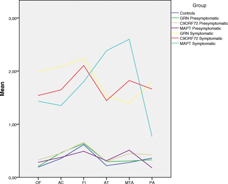
In patients with frontotemporal dementia, it has been shown that brain atrophy occurs earliest in the anterior cingulate, insula and frontal lobes. We used visual rating scales to investigate whether identifying atrophy in these areas may be helpful in distinguishing symptomatic patients carrying different causal mutations in the microtubule-associated protein tau (MAPT), progranulin (GRN) and chromosome 9 open reading frame (C9ORF72) genes. We also analysed asymptomatic carriers to see whether it was possible to visually identify brain atrophy before the appearance of symptoms.
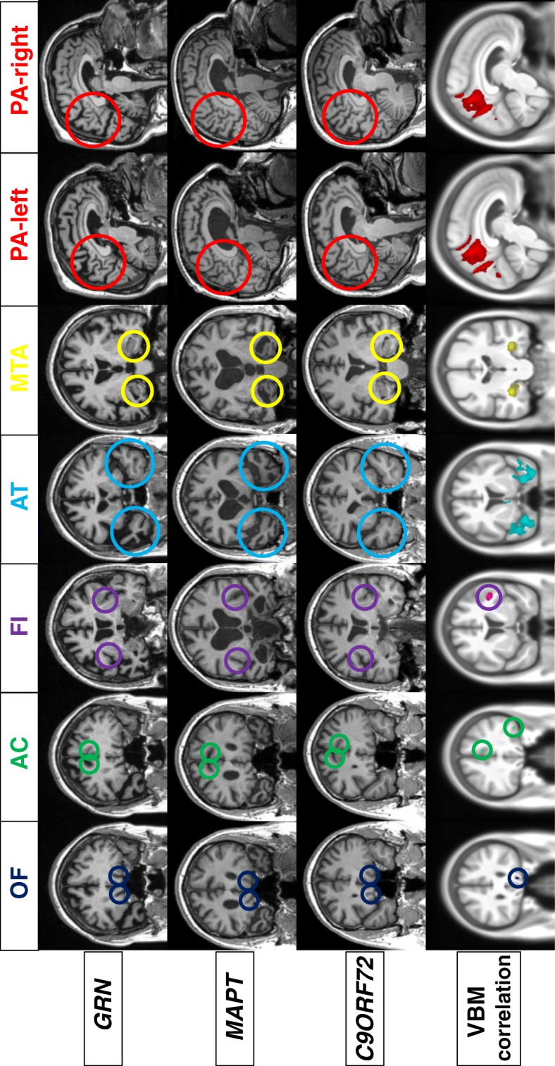
Magnetic resonance imaging of 343 subjects (63 symptomatic mutation carriers, 132 presymptomatic mutation carriers and 148 control subjects) from the Genetic Frontotemporal Dementia Initiative study were analysed by two trained raters using a protocol of six visual rating scales that identified atrophy in key regions of the brain (orbitofrontal, anterior cingulate, frontoinsula, anterior and medial temporal lobes and posterior cortical areas).
Intra- and interrater agreement were greater than 0.73 for all the scales. Voxel-based morphometric analysis demonstrated a strong correlation between the visual rating scale scores and grey matter atrophy in the same region for each of the scales. Typical patterns of atrophy were identified: symmetric anterior and medial temporal lobe involvement for MAPT, asymmetric frontal and parietal loss for GRN, and a more widespread pattern for C9ORF72. Presymptomatic MAPT carriers showed greater atrophy in the medial temporal region than control subjects, but the visual rating scales could not identify presymptomatic atrophy in GRN or C9ORF72 carriers.
These simple-to-use and reproducible scales may be useful tools in the clinical setting for the discrimination of different mutations of frontotemporal dementia, and they may even help to identify atrophy prior to onset in those with MAPT mutations.
在额颞叶痴呆患者中,已经表明大脑萎缩最早发生在前扣带回、脑岛和额叶。我们使用视觉评分量表来研究识别这些区域的萎缩是否有助于区分携带微管相关蛋白 tau(MAPT)、颗粒蛋白前体(GRN)和 9 号染色体开放阅读框(C9ORF72)基因突变的有症状患者。我们还分析了无症状携带者,以观察是否有可能在出现症状之前通过视觉识别脑萎缩。
对来自遗传额颞叶痴呆倡议研究的 343 名受试者(63 名有症状突变携带者、132 名无症状突变携带者和 148 名对照受试者)的磁共振成像进行了分析,由两名经过培训的评分员使用 6 种视觉评分量表的方案进行分析,这些量表确定了大脑关键区域(眶额、前扣带回、额岛、前颞叶和内侧颞叶以及皮质后区)的萎缩。
所有量表的组内和组间一致性均大于 0.73。基于体素的形态计量学分析表明,在每个量表中,视觉评分量表评分与同一区域的灰质萎缩之间存在很强的相关性。确定了典型的萎缩模式:MAPT 为对称的前内侧颞叶受累,GRN 为不对称的额顶叶丢失,C9ORF72 为更广泛的模式。无症状的 MAPT 携带者的内侧颞叶区域比对照组有更大的萎缩,但视觉评分量表无法识别 GRN 或 C9ORF72 携带者的无症状萎缩。
这些易于使用且可重复的量表可能是临床环境中区分不同额颞叶痴呆突变的有用工具,它们甚至可能有助于在携带 MAPT 突变的患者中识别发病前的萎缩。