Otter Simon, Payne Catherine, Jones Anna-Marie, Webborn Nick, Watt Peter
School of Health Sciences, University of Brighton, 49 Darley Rd, Eastbourne, BN20 7UR, UK.
Centre for Regenerative Medicine and Devices, University of Brighton, Lewes Road, Brighton, BN2 4AT, UK.
BMC Musculoskelet Disord. 2020 Oct 7;21(1):658. doi: 10.1186/s12891-020-03598-3.
Gout has been associated with weaker foot/leg muscles and altered gait patterns. There is also evidence of on-going foot pain and an increased risk of tendinopathy, with the Achilles and patella tendons most frequently affected in gout. Additionally, the inflammation associated with gout may change tissue elasticity. Ultrasound imaging utilising shear wave elastography (SWE) offers a non-invasive method of quantifying changes in tendon stiffness. SWE findings have not previously been reported in individuals with gout. We sought to determine differences in Achilles tendon stiffness in people with gout compared to controls (non-gout).
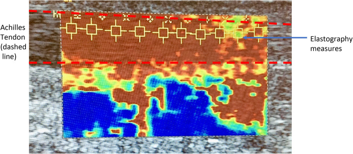
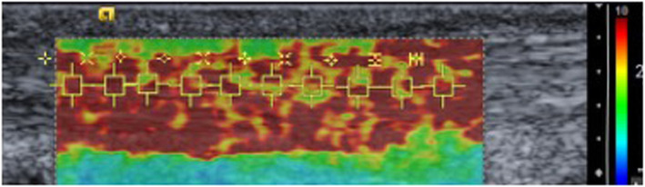
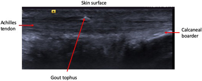
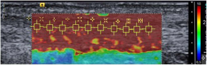
A cross sectional study comparing 24 people with gout and 26 age/sex-matched controls. Clinical and demographic data were collated, and US imaging used to determine tendon thickness, presence of gouty tophi and/or aggregates and levels of angiogenesis. Ten shear wave elastography (SWE) measures were taken along the centre of a longitudinal section of the mid-portion of each Achilles tendon. Prior to data collection, intra-observer error was good (>0.69). Data were summarised using descriptive statistics and a repeated measures ANCOVA was used to compare SWE measures between the two groups for the left and right foot separately after accounting for Body Mass Index (BMI).
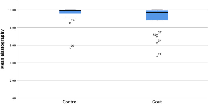
A small proportion of those with gout presented with intra-tendon aggregates and/or intra-tendon tophi in one or both tendons. There was no statistically significant difference in tendon thickness between groups. Neo-vascularity was present in a third of gout participants. SWE findings demonstrated significantly reduced tendon stiffness in those with gout compared to controls: right Achilles mdiff =1.04 m/s (95% CI (0.38 to 1.7) p = 0.003 and left Achilles mdiff = 0.7 m/s (95% CI 0.09 to 1.32) p = 0.025. No relationship between the presence of tophi and SWE values were detected.
Subjects with chronic gout show significantly reduced Achilles tendon stiffness compared to non-gout controls. From a clinical standpoint, our findings were similar to SWE measurements in subjects with Achilles tendinopathy and who did not have gout.
痛风与足部/腿部肌肉较弱及步态模式改变有关。也有证据表明存在持续性足部疼痛以及肌腱病风险增加,痛风患者中最常受累的是跟腱和髌腱。此外,与痛风相关的炎症可能会改变组织弹性。利用剪切波弹性成像(SWE)的超声成像提供了一种量化肌腱硬度变化的非侵入性方法。此前尚未有痛风患者的SWE检查结果报告。我们试图确定痛风患者与对照组(非痛风患者)跟腱硬度的差异。
一项横断面研究,比较了24名痛风患者和26名年龄/性别匹配的对照组。整理了临床和人口统计学数据,并使用超声成像来确定肌腱厚度、痛风石和/或聚集体的存在情况以及血管生成水平。在每条跟腱中部纵向切面的中心进行10次剪切波弹性成像(SWE)测量。在数据收集之前,观察者内误差良好(>0.69)。数据采用描述性统计进行汇总,并在考虑体重指数(BMI)后,使用重复测量协方差分析分别比较两组左右脚的SWE测量值。
一小部分痛风患者的一条或两条肌腱出现肌腱内聚集体和/或肌腱内痛风石。两组之间肌腱厚度无统计学显著差异。三分之一的痛风参与者存在新生血管。SWE检查结果显示,与对照组相比,痛风患者的肌腱硬度显著降低:右侧跟腱mdiff = 1.04 m/s(95%CI(0.38至1.7),p = 0.003;左侧跟腱mdiff = 0.7 m/s(95%CI 0.09至1.32),p = 0.025。未检测到痛风石的存在与SWE值之间的关系。
与非痛风对照组相比,慢性痛风患者的跟腱硬度显著降低。从临床角度来看,我们的研究结果与跟腱病但无痛风患者的SWE测量结果相似。