Laboratório de Pesquisa em Apicomplexa, Instituto Carlos Chagas, Fiocruz Paraná, Curitiba, PR, Brazil.
Laboratório de Doenças Tropicais Prof. Luiz Jacintho da Silva, Departamento de Genética, Evolução, Microbiologia e Imunologia, Instituto de Biologia, Universidade Estadual de Campinas (UNICAMP), Campinas, SP, Brazil.
Sci Rep. 2020 Oct 7;10(1):16706. doi: 10.1038/s41598-020-73713-w.
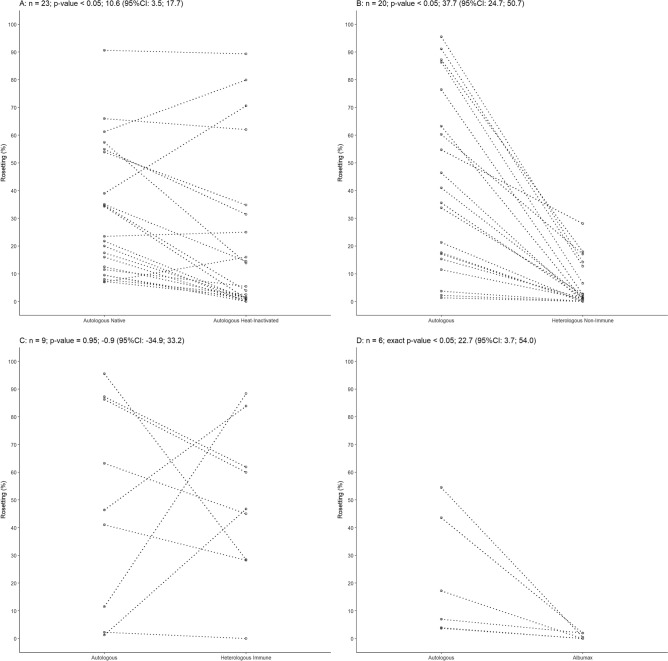
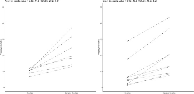
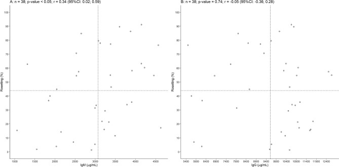
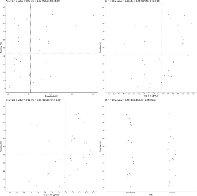
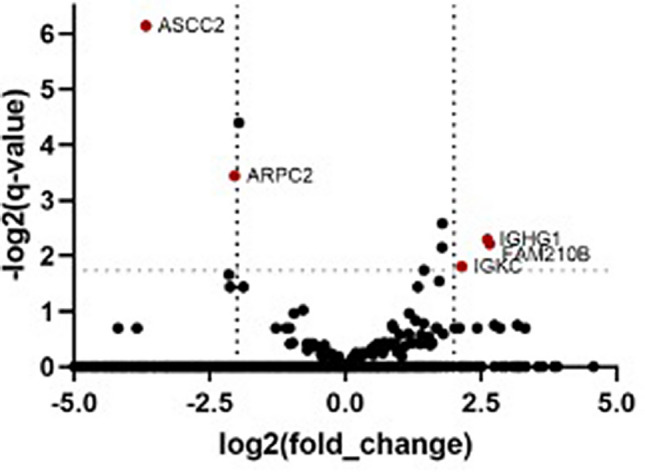
Plasmodium vivax is the most prevalent cause of malaria outside of Africa. P. vivax biology and pathogenesis are still poorly understood. The role of one highly occurring phenotype in particular where infected reticulocytes cytoadhere to noninfected normocytes, forming rosettes, remains unknown. Here, using a range of ex vivo approaches, we showed that P. vivax rosetting rates were enhanced by plasma of infected patients and that total immunoglobulin M levels correlated with rosetting frequency. Moreover, rosetting rates were also correlated with parasitemia, IL-6 and IL-10 levels in infected patients. Transcriptomic analysis of peripheral leukocytes from P. vivax-infected patients with low or moderated rosetting rates identified differentially expressed genes related to human host phagocytosis pathway. In addition, phagocytosis assay showed that rosetting parasites were less phagocyted. Collectively, these results showed that rosette formation plays a role in host immune response by hampering leukocyte phagocytosis. Thus, these findings suggest that rosetting could be an effective P. vivax immune evasion strategy.
卵形疟原虫是非洲以外地区最常见的疟疾病原体。然而,人们对卵形疟原虫的生物学和发病机制仍了解甚少。其中一个高度发生的表型尤其值得关注,即感染的网织红细胞黏附到未感染的正常红细胞上,形成玫瑰花结。在这里,我们使用一系列的离体方法表明,感染患者的血浆可增强卵形疟原虫的玫瑰花结形成率,且总免疫球蛋白 M 水平与玫瑰花结形成频率相关。此外,玫瑰花结形成率还与感染患者的寄生虫血症、IL-6 和 IL-10 水平相关。对低或中度玫瑰花结形成率的卵形疟原虫感染患者外周血白细胞进行转录组分析,发现了与人类宿主吞噬途径相关的差异表达基因。此外,吞噬试验表明,玫瑰花结形成的寄生虫更不易被吞噬。总之,这些结果表明,玫瑰花结的形成通过阻碍白细胞吞噬作用,在宿主免疫反应中发挥作用。因此,这些发现表明,玫瑰花结可能是卵形疟原虫有效的免疫逃避策略。