• 文献检索
  • 文档翻译
  • 深度研究
  • 学术资讯
  • Suppr Zotero 插件Zotero 插件
  • 邀请有礼
  • 套餐&价格
  • 历史记录
应用&插件
Suppr Zotero 插件Zotero 插件浏览器插件Mac 客户端Windows 客户端微信小程序
定价
高级版会员购买积分包购买API积分包
服务
文献检索文档翻译深度研究API 文档MCP 服务
关于我们
关于 Suppr公司介绍联系我们用户协议隐私条款
关注我们

Suppr 超能文献

核心技术专利:CN118964589B侵权必究
粤ICP备2023148730 号-1Suppr @ 2026

文献检索

告别复杂PubMed语法,用中文像聊天一样搜索,搜遍4000万医学文献。AI智能推荐,让科研检索更轻松。

立即免费搜索

文件翻译

保留排版,准确专业,支持PDF/Word/PPT等文件格式,支持 12+语言互译。

免费翻译文档

深度研究

AI帮你快速写综述,25分钟生成高质量综述,智能提取关键信息,辅助科研写作。

立即免费体验

荧光可视化引导手术对口腔鳞状细胞癌局部复发的影响:一项随机临床试验。

Effect of Fluorescence Visualization-Guided Surgery on Local Recurrence of Oral Squamous Cell Carcinoma: A Randomized Clinical Trial.

机构信息

Department of Surgery, Faculty of Medicine, The University of British Columbia, Vancouver, British Columbia, Canada.

Department of Statistics, Faculty of Science, The University of British Columbia, Vancouver, British Columbia, Canada.

出版信息

JAMA Otolaryngol Head Neck Surg. 2020 Dec 1;146(12):1149-1155. doi: 10.1001/jamaoto.2020.3147.

DOI:10.1001/jamaoto.2020.3147
PMID:33034628
原文链接:https://pmc.ncbi.nlm.nih.gov/articles/PMC7545352/
Abstract

IMPORTANCE

High local recurrence rates with aggressive disease remain the main concern in oral cancer survival. Use of a translational device using fluorescence visualization (FV) approved by the US Food and Drug Administration and Health Canada, has shown a marked reduction in the 3-year local recurrence rate of high-grade oral lesions in a single-center observational study.

OBJECTIVE

To determine whether FV- guided surgery can improve local control rates in the treatment of in situ or T1 to T2 category oral squamous cell carcinoma (OSCC).

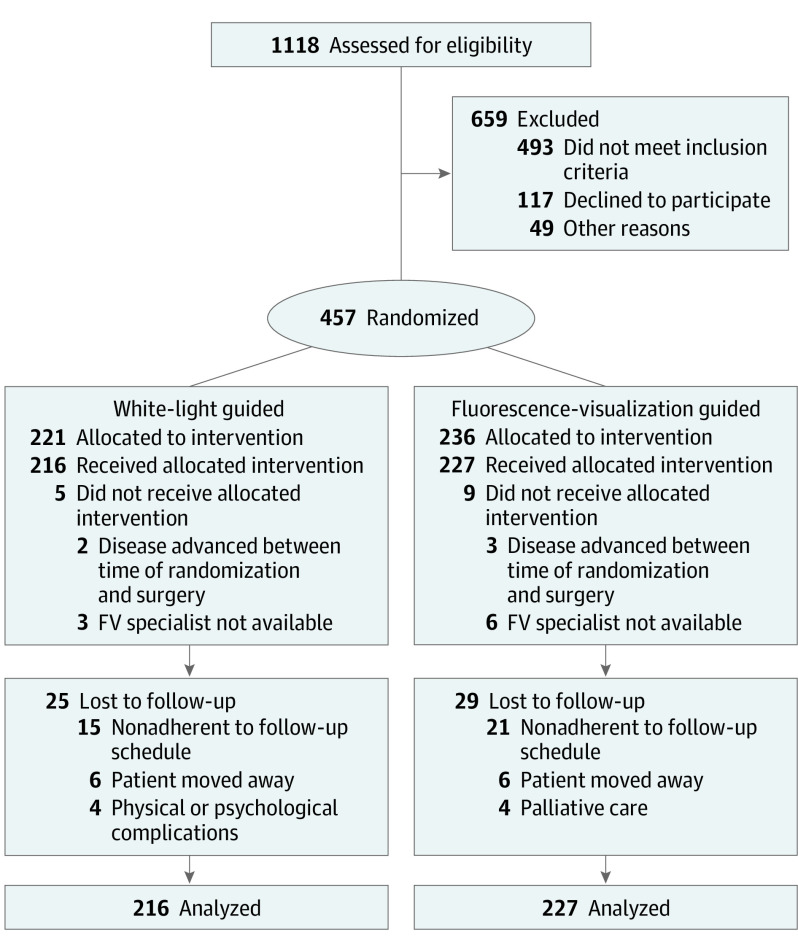
DESIGN, SETTING, AND PARTICIPANTS: A multicenter randomized clinical trial was conducted in a surgical setting. A total of 457 patients were enrolled between January 18, 2010, and April 30, 2015. Data analysis of the intention-to-treat population was performed from April 3, 2019, to March 20, 2020. Patients with histologically confirmed high-grade dysplasia/carcinoma in situ or T1 to T2 category OSCC were randomized to receive traditional peroral surgery or FV-guided surgery.

INTERVENTION

Fluorescence visualization during surgery.

MAIN OUTCOMES AND MEASURES

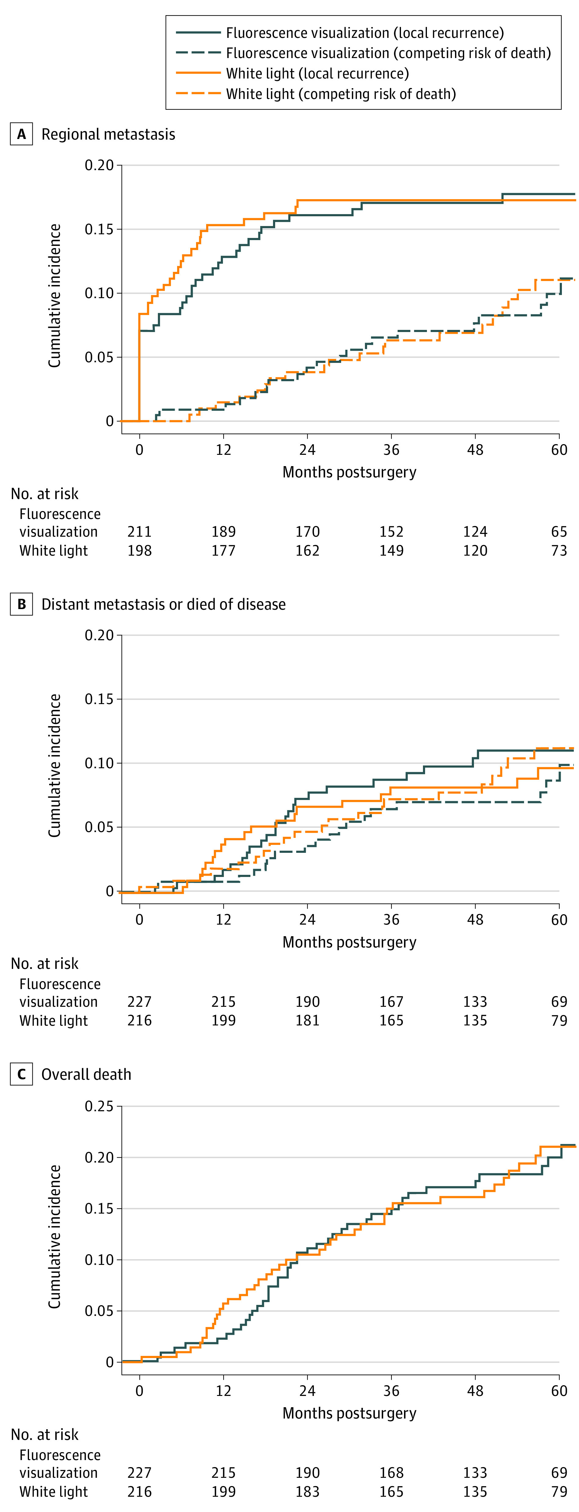
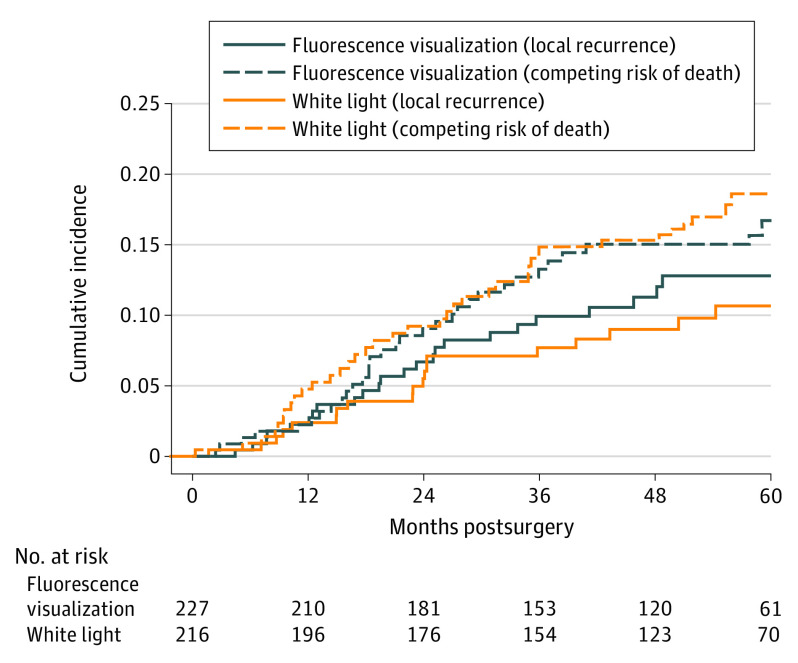
The primary outcome was local recurrence of OSCC. Secondary outcomes were failure of the first-pass margin, defined as a histologically confirmed positive margin for severe dysplasia or greater histologic change of the main specimen (ie, not the margins taken from the resection bed), regional or distant metastasis, and death due to disease.

RESULTS

Of the 457 patients enrolled in the study, 443 patients (264 [59.6%] men; mean [SD] age, 61.5 [13.3] years) completed the randomized treatment: 227 FV-guided and 216 non-FV guided surgery. The median follow-up was 52 (range, 0.29-90.8) months. In total, 45 patients (10.2%) experienced local recurrence. The 3-year local recurrence rate was 9.4% in the FV-guided group and 7.2% in the non-FV group (difference, 2.2%; 95% CI, -3.2% to 7.4%). Other similarities between the FV vs non-FV groups included failure of first-pass margin (68/227 [30.0%]) vs 65/216 [30.1%]), regional failure (39/227 [17.2%] vs 37/216 [17.1%]), disease-specific survival (23/227 [10.1%] vs 19/26 [8.8%]), and overall survival (41/227 [18.1%] vs 38/216 [17.6%]) were also similar between groups. No adverse events were judged to be related to the intervention.

CONCLUSIONS AND RELEVANCE

In this randomized clinical trial, FV-guided surgery did not improve local control rates in the treatment of patients with in situ or T1 to T2 category oral cancer. Under a controlled environment, FV-guided surgery did not have an evident effect in reduction of local recurrence for localized OSCC. This result suggests that attention be directed to strategies other than improving definitions of nonapparent disease at clinical margins to identify the sources of local recurrence.

TRIAL REGISTRATION

ClinicalTrial.gov Identifier: NCT01039298.

摘要

重要性:在口腔癌的生存中,高局部复发率和侵袭性疾病仍然是主要关注点。使用美国食品和药物管理局和加拿大卫生部批准的翻译设备(荧光可视化[FV]),在一项单中心观察性研究中,已显示出高级别口腔病变的 3 年局部复发率显著降低。

目的:确定 FV 引导手术是否可以改善原位或 T1 至 T2 类口腔鳞状细胞癌(OSCC)的局部控制率。

设计、地点和参与者:在手术环境中进行了一项多中心随机临床试验。2010 年 1 月 18 日至 2015 年 4 月 30 日期间共纳入 457 例患者。对意向治疗人群进行数据分析的时间为 2019 年 4 月 3 日至 2020 年 3 月 20 日。将组织学证实为高级别发育不良/原位癌或 T1 至 T2 类 OSCC 的患者随机分为接受传统经口手术或 FV 引导手术。

干预措施:手术过程中的荧光可视化。

主要结果和措施:主要结局是 OSCC 的局部复发。次要结局是首次通过边缘失败,定义为主要标本的组织学证实阳性边缘(即不是从切除床获得的边缘),区域或远处转移,以及疾病相关死亡。

结果:在这项研究中,共纳入 457 例患者,443 例患者(264 [59.6%] 名男性;平均[SD]年龄 61.5 [13.3] 岁)完成了随机治疗:227 例 FV 引导和 216 例非 FV 引导手术。中位随访时间为 52 个月(范围,0.29-90.8)。共有 45 例(10.2%)患者出现局部复发。FV 引导组的 3 年局部复发率为 9.4%,非 FV 组为 7.2%(差异 2.2%;95%CI,-3.2%至 7.4%)。FV 与非 FV 组之间的其他相似之处还包括首次通过边缘失败(68/227 [30.0%])与 65/216 [30.1%])、区域失败(39/227 [17.2%])与 37/216 [17.1%])、疾病特异性生存率(23/227 [10.1%])与 19/26 [8.8%])和总生存率(41/227 [18.1%])也相似。没有不良事件被认为与干预有关。

结论和相关性:在这项随机临床试验中,FV 引导手术并未提高原位或 T1 至 T2 类口腔癌患者的局部控制率。在受控环境下,FV 引导手术在降低局部复发率方面对局限性 OSCC 没有明显作用。这一结果表明,需要关注的是在临床边缘改善非明显疾病的定义之外的策略,以确定局部复发的来源。

试验注册:ClinicalTrials.gov 标识符:NCT01039298。

https://cdn.ncbi.nlm.nih.gov/pmc/blobs/3cf1/7545352/eb896c3d26c4/jamaotolaryngolheadnecksurg-e203147-g003.jpg
https://cdn.ncbi.nlm.nih.gov/pmc/blobs/3cf1/7545352/49de296ca032/jamaotolaryngolheadnecksurg-e203147-g001.jpg
https://cdn.ncbi.nlm.nih.gov/pmc/blobs/3cf1/7545352/92acfd13c998/jamaotolaryngolheadnecksurg-e203147-g002.jpg
https://cdn.ncbi.nlm.nih.gov/pmc/blobs/3cf1/7545352/eb896c3d26c4/jamaotolaryngolheadnecksurg-e203147-g003.jpg

相似文献

1
Effect of Fluorescence Visualization-Guided Surgery on Local Recurrence of Oral Squamous Cell Carcinoma: A Randomized Clinical Trial.荧光可视化引导手术对口腔鳞状细胞癌局部复发的影响:一项随机临床试验。
JAMA Otolaryngol Head Neck Surg. 2020 Dec 1;146(12):1149-1155. doi: 10.1001/jamaoto.2020.3147.
2
Fluorescence Visualization-Guided Surgery for Early-Stage Oral Cancer.荧光可视化引导的早期口腔癌手术
JAMA Otolaryngol Head Neck Surg. 2016 Mar;142(3):209-16. doi: 10.1001/jamaoto.2015.3211.
3
Canadian Optically-guided approach for Oral Lesions Surgical (COOLS) trial: study protocol for a randomized controlled trial.加拿大光学引导口腔病变手术(COOLS)试验:一项随机对照试验的研究方案。
BMC Cancer. 2011 Oct 25;11:462. doi: 10.1186/1471-2407-11-462.
4
Clinicopathological parameters, recurrence, locoregional and distant metastasis in 115 T1-T2 oral squamous cell carcinoma patients.115例T1-T2期口腔鳞状细胞癌患者的临床病理参数、复发情况、局部区域及远处转移情况
Head Neck Oncol. 2010 Apr 20;2:9. doi: 10.1186/1758-3284-2-9.
5
Association of Main Specimen and Tumor Bed Margin Status With Local Recurrence and Survival in Oral Cancer Surgery.口腔癌手术中主标本和肿瘤床切缘状态与局部复发和生存的关系。
JAMA Otolaryngol Head Neck Surg. 2016 Dec 1;142(12):1191-1198. doi: 10.1001/jamaoto.2016.2329.
6
Margin Assessment Methods in Oral Cavity Squamous Cell Carcinoma and Recurrence: Tumor Bed vs Resection Specimen Sampling.口腔鳞状细胞癌及复发的边缘评估方法:肿瘤床与切除标本取样。
JAMA Otolaryngol Head Neck Surg. 2023 Nov 1;149(11):1011-1020. doi: 10.1001/jamaoto.2023.2982.
7
Prospective evaluation of outcome after transoral CO(2) laser resection of T1/T2 oral squamous cell carcinoma.经口二氧化碳激光切除T1/T2期口腔鳞状细胞癌术后结局的前瞻性评估
Oral Surg Oral Med Oral Pathol Oral Radiol Endod. 2011 Aug;112(2):180-7. doi: 10.1016/j.tripleo.2010.09.078. Epub 2011 Jan 12.
8
Impact of dysplastic surgical margins for patients with oral squamous cell carcinoma.口腔鳞状细胞癌患者的手术不典型边缘的影响。
Oral Oncol. 2019 Oct;97:1-6. doi: 10.1016/j.oraloncology.2019.07.015. Epub 2019 Jul 31.
9
Comprehensive survival analysis of oral squamous cell carcinoma patients undergoing initial radical surgery.口腔鳞状细胞癌患者初始根治性手术的综合生存分析。
BMC Oral Health. 2024 Aug 9;24(1):919. doi: 10.1186/s12903-024-04690-z.
10
The relevance of surgical margins in clinically early oral squamous cell carcinoma.手术切缘在临床早期口腔鳞状细胞癌中的相关性。
Oral Oncol. 2020 Nov;110:104913. doi: 10.1016/j.oraloncology.2020.104913. Epub 2020 Jul 22.

引用本文的文献

1
Salivary cell-free DNA methylation analysis for oncological monitoring of surgical resection of oral squamous cell carcinoma.用于口腔鳞状细胞癌手术切除肿瘤学监测的唾液游离DNA甲基化分析
Front Oral Health. 2025 Jun 11;6:1614371. doi: 10.3389/froh.2025.1614371. eCollection 2025.
2
Fluorescence Imaging-Assessed Surgical Margin Detection in Head and Neck Oncology by Passive and Active Targeting.通过被动和主动靶向的荧光成像评估头颈部肿瘤手术切缘
Mol Diagn Ther. 2025 May 8. doi: 10.1007/s40291-025-00781-x.
3
Recent advances of photodiagnosis and treatment for head and neck squamous cell carcinoma.

本文引用的文献

1
Fluorescence Visualization-Guided Surgery for Early-Stage Oral Cancer.荧光可视化引导的早期口腔癌手术
JAMA Otolaryngol Head Neck Surg. 2016 Mar;142(3):209-16. doi: 10.1001/jamaoto.2015.3211.
2
Cancer incidence and mortality worldwide: sources, methods and major patterns in GLOBOCAN 2012.全球癌症发病与死亡:GLOBOCAN 2012 数据源、方法与主要模式。
Int J Cancer. 2015 Mar 1;136(5):E359-86. doi: 10.1002/ijc.29210. Epub 2014 Oct 9.
3
Canadian Optically-guided approach for Oral Lesions Surgical (COOLS) trial: study protocol for a randomized controlled trial.
头颈部鳞状细胞癌的光诊断与治疗的最新进展
Neoplasia. 2025 Feb;60:101118. doi: 10.1016/j.neo.2024.101118. Epub 2024 Dec 24.
4
Feasibility of an MR-based digital specimen for tongue cancer resection specimens: a novel approach for margin evaluation.基于磁共振成像的舌癌切除标本数字标本的可行性:一种切缘评估的新方法。
Front Oncol. 2024 Mar 28;14:1342857. doi: 10.3389/fonc.2024.1342857. eCollection 2024.
5
Intraoperative Techniques That Define the Mucosal Margins of Oral Cancer In-Vivo: A Systematic Review.确定口腔癌体内黏膜边缘的术中技术:一项系统综述。
Cancers (Basel). 2024 Mar 14;16(6):1148. doi: 10.3390/cancers16061148.
6
A Prospective Double-Blinded Comparison of Reflectance Confocal Microscopy with Conventional Histopathology for In Vivo Assessment in Oral Cancer.在口腔癌的体内评估中,反射共聚焦显微镜与常规组织病理学的前瞻性双盲比较。
Clin Cancer Res. 2024 Jun 3;30(11):2486-2496. doi: 10.1158/1078-0432.CCR-23-1361.
7
Clinical Experience with Autofluorescence Guided Oral Squamous Cell Carcinoma Surgery.自体荧光引导下口腔鳞状细胞癌手术的临床经验
Diagnostics (Basel). 2023 Oct 10;13(20):3161. doi: 10.3390/diagnostics13203161.
8
Interventions for the treatment of oral cavity and oropharyngeal cancers: surgical treatment.口腔和口咽癌的治疗干预措施:手术治疗。
Cochrane Database Syst Rev. 2023 Aug 31;8(8):CD006205. doi: 10.1002/14651858.CD006205.pub5.
9
Trends in Positive Surgical Margins in cT3-T4 Oral Cavity Squamous Cell Carcinoma.cT3-T4 期口腔鳞癌阳性切缘趋势。
Otolaryngol Head Neck Surg. 2023 Nov;169(5):1200-1207. doi: 10.1002/ohn.377. Epub 2023 May 26.
10
Anatomy-Specific Classification Model Using Label-Free FLIm to Aid Intraoperative Surgical Guidance of Head and Neck Cancer.基于无标记荧光寿命成像的解剖特异性分类模型辅助头颈部癌症术中手术指导
IEEE Trans Biomed Eng. 2023 Oct;70(10):2863-2873. doi: 10.1109/TBME.2023.3266678. Epub 2023 Sep 27.
加拿大光学引导口腔病变手术(COOLS)试验:一项随机对照试验的研究方案。
BMC Cancer. 2011 Oct 25;11:462. doi: 10.1186/1471-2407-11-462.
4
Potential use of quantitative tissue phenotype to predict malignant risk for oral premalignant lesions.定量组织表型在预测口腔癌前病变恶性风险中的潜在应用。
Cancer Res. 2008 May 1;68(9):3099-107. doi: 10.1158/0008-5472.CAN-07-2113.
5
Speech Handicap Index in patients with oral and pharyngeal cancer: better understanding of patients' complaints.口腔和咽癌患者的言语障碍指数:更好地理解患者的主诉。
Head Neck. 2008 Jul;30(7):868-74. doi: 10.1002/hed.20795.
6
Multiple cancer prevalence: a growing challenge in long-term survivorship.多种癌症患病率:长期生存中日益严峻的挑战。
Cancer Epidemiol Biomarkers Prev. 2007 Mar;16(3):566-71. doi: 10.1158/1055-9965.EPI-06-0782.
7
Simple device for the direct visualization of oral-cavity tissue fluorescence.用于直接观察口腔组织荧光的简易装置。
J Biomed Opt. 2006 Mar-Apr;11(2):024006. doi: 10.1117/1.2193157.
8
US valuation of the EQ-5D health states: development and testing of the D1 valuation model.美国EQ-5D健康状态的估值:D1估值模型的开发与测试
Med Care. 2005 Mar;43(3):203-20. doi: 10.1097/00005650-200503000-00003.
9
Field cancerization in oral stratified squamous epithelium; clinical implications of multicentric origin.口腔复层鳞状上皮中的场癌化;多中心起源的临床意义。
Cancer. 1953 Sep;6(5):963-8. doi: 10.1002/1097-0142(195309)6:5<963::aid-cncr2820060515>3.0.co;2-q.
10
A genetic explanation of Slaughter's concept of field cancerization: evidence and clinical implications.斯劳特场癌化概念的遗传学解释:证据及临床意义
Cancer Res. 2003 Apr 15;63(8):1727-30.