• 文献检索
  • 文档翻译
  • 深度研究
  • 学术资讯
  • Suppr Zotero 插件Zotero 插件
  • 邀请有礼
  • 套餐&价格
  • 历史记录
应用&插件
Suppr Zotero 插件Zotero 插件浏览器插件Mac 客户端Windows 客户端微信小程序
定价
高级版会员购买积分包购买API积分包
服务
文献检索文档翻译深度研究API 文档MCP 服务
关于我们
关于 Suppr公司介绍联系我们用户协议隐私条款
关注我们

Suppr 超能文献

核心技术专利:CN118964589B侵权必究
粤ICP备2023148730 号-1Suppr @ 2026

文献检索

告别复杂PubMed语法,用中文像聊天一样搜索,搜遍4000万医学文献。AI智能推荐,让科研检索更轻松。

立即免费搜索

文件翻译

保留排版,准确专业,支持PDF/Word/PPT等文件格式,支持 12+语言互译。

免费翻译文档

深度研究

AI帮你快速写综述,25分钟生成高质量综述,智能提取关键信息,辅助科研写作。

立即免费体验

健康猕猴大脑皮质中扩散峰度测量的神经解剖学基础

Neuroanatomical underpinning of diffusion kurtosis measurements in the cerebral cortex of healthy macaque brains.

作者信息

Zhu Tianjia, Peng Qinmu, Ouyang Austin, Huang Hao

机构信息

Department of Radiology, Children's Hospital of Philadelphia, Philadelphia, Pennsylvania, USA.

Department of Bioengineering, University of Pennsylvania, Philadelphia, Pennsylvania, USA.

出版信息

Magn Reson Med. 2021 Apr;85(4):1895-1908. doi: 10.1002/mrm.28548. Epub 2020 Oct 15.

DOI:10.1002/mrm.28548
PMID:33058286
原文链接:https://pmc.ncbi.nlm.nih.gov/articles/PMC8934732/
Abstract

PURPOSE

To investigate the neuroanatomical underpinning of healthy macaque brain cortical microstructure measured by diffusion kurtosis imaging (DKI), which characterizes non-Gaussian water diffusion.

METHODS

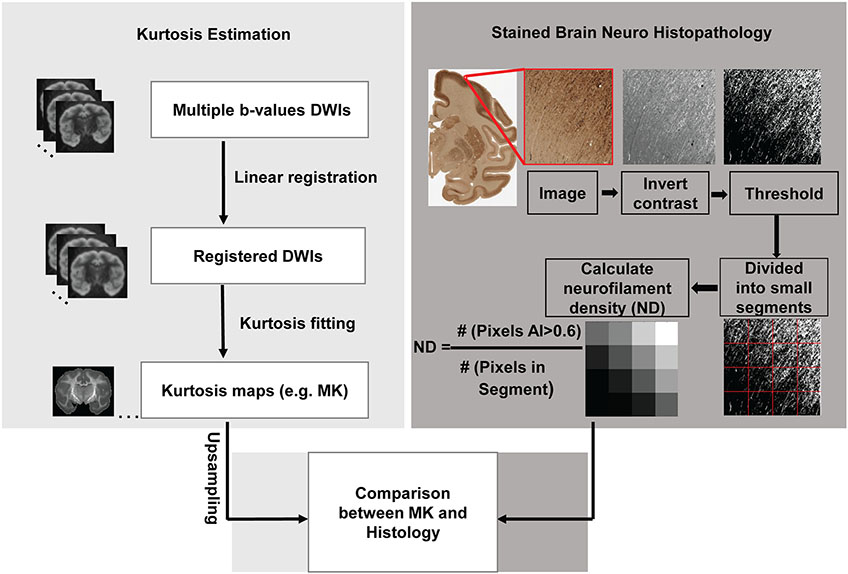
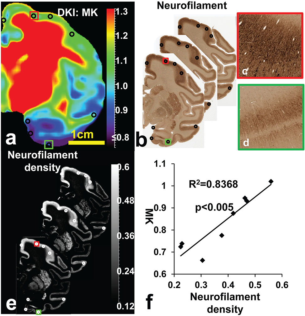
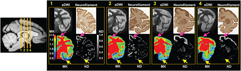
High-resolution DKI was acquired from 6 postmortem macaque brains. Neurofilament density (ND) was quantified based on structure tensor from neurofilament histological images of a different macaque brain sample. After alignment of DKI-derived mean kurtosis (MK) maps to the histological images, MK and histology-based ND were measured at corresponding regions of interests characterized by distinguished cortical MK values in the prefrontal/precentral-postcentral and temporal cortices. Pearson correlation was performed to test significant correlation between these cortical MK and ND measurements.

RESULTS

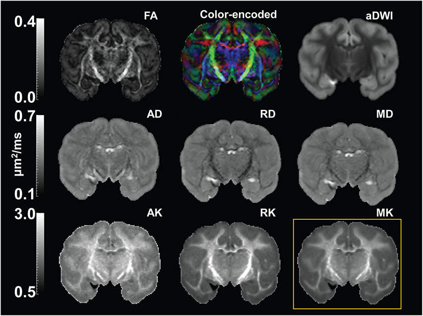
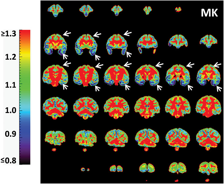
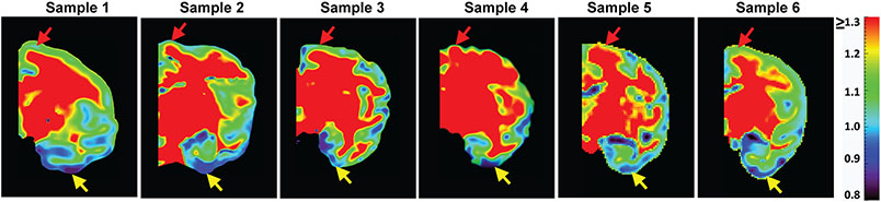
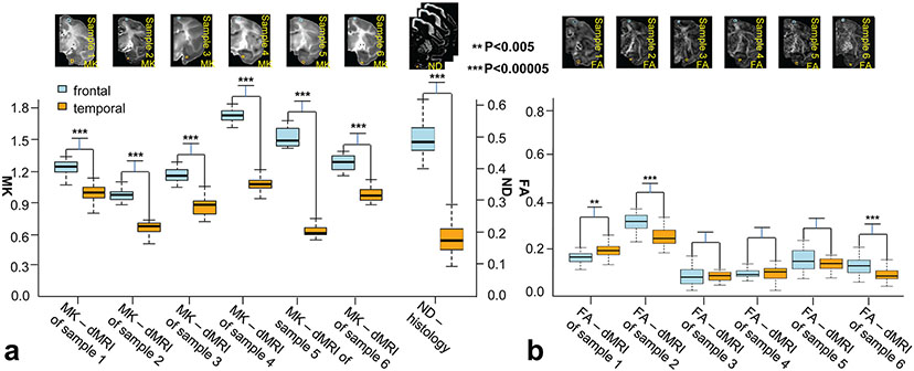
Heterogeneity of cortical MK across different cortical regions was revealed, with significantly and consistently higher MK measurements in the prefrontal/precentral-postcentral cortex compared to those in the temporal cortex across all six scanned macaque brains. Corresponding higher ND measurements in the prefrontal/precentral-postcentral cortex than in the temporal cortex were also found. The heterogeneity of cortical MK is associated with heterogeneity of histology-based ND measurements, with significant correlation between cortical MK and corresponding ND measurements (P < .005).

CONCLUSION

These findings suggested that DKI-derived MK can potentially be an effective noninvasive biomarker quantifying underlying neuroanatomical complexity inside the cerebral cortical mantle for clinical and neuroscientific research.

摘要

目的

研究通过扩散峰度成像(DKI)测量的健康猕猴脑皮质微观结构的神经解剖学基础,DKI可表征非高斯水扩散。

方法

从6只猕猴死后大脑获取高分辨率DKI。基于来自另一只猕猴脑样本的神经丝组织学图像的结构张量对神经丝密度(ND)进行量化。在将DKI衍生的平均峰度(MK)图与组织学图像对齐后,在额叶/中央前回-中央后回和颞叶皮质中以不同的皮质MK值为特征的相应感兴趣区域测量MK和基于组织学的ND。进行Pearson相关性分析以检验这些皮质MK和ND测量值之间的显著相关性。

结果

揭示了不同皮质区域皮质MK的异质性,在所有六只扫描的猕猴脑中,额叶/中央前回-中央后回皮质中的MK测量值明显且始终高于颞叶皮质。还发现额叶/中央前回-中央后回皮质中的ND测量值相应高于颞叶皮质。皮质MK的异质性与基于组织学的ND测量值的异质性相关,皮质MK与相应的ND测量值之间存在显著相关性(P <.005)。

结论

这些发现表明,DKI衍生的MK可能潜在地成为一种有效的非侵入性生物标志物,用于量化临床和神经科学研究中大脑皮质层内潜在的神经解剖学复杂性。

https://cdn.ncbi.nlm.nih.gov/pmc/blobs/43e7/8934732/0b2a69a547e4/nihms-1784791-f0007.jpg
https://cdn.ncbi.nlm.nih.gov/pmc/blobs/43e7/8934732/df5d578945d4/nihms-1784791-f0001.jpg
https://cdn.ncbi.nlm.nih.gov/pmc/blobs/43e7/8934732/e7ff05f03549/nihms-1784791-f0002.jpg
https://cdn.ncbi.nlm.nih.gov/pmc/blobs/43e7/8934732/5e0fe97503e6/nihms-1784791-f0003.jpg
https://cdn.ncbi.nlm.nih.gov/pmc/blobs/43e7/8934732/aefee8d504a0/nihms-1784791-f0004.jpg
https://cdn.ncbi.nlm.nih.gov/pmc/blobs/43e7/8934732/8f7ba282152f/nihms-1784791-f0005.jpg
https://cdn.ncbi.nlm.nih.gov/pmc/blobs/43e7/8934732/a8011ded0fde/nihms-1784791-f0006.jpg
https://cdn.ncbi.nlm.nih.gov/pmc/blobs/43e7/8934732/0b2a69a547e4/nihms-1784791-f0007.jpg

相似文献

1
Neuroanatomical underpinning of diffusion kurtosis measurements in the cerebral cortex of healthy macaque brains.健康猕猴大脑皮质中扩散峰度测量的神经解剖学基础
Magn Reson Med. 2021 Apr;85(4):1895-1908. doi: 10.1002/mrm.28548. Epub 2020 Oct 15.
2
Differential cortical microstructural maturation in the preterm human brain with diffusion kurtosis and tensor imaging.应用扩散峰度和张量成像技术对早产儿大脑皮质微观结构成熟度的差异研究。
Proc Natl Acad Sci U S A. 2019 Mar 5;116(10):4681-4688. doi: 10.1073/pnas.1812156116. Epub 2019 Feb 19.
3
Diffusion kurtosis imaging of gray matter in schizophrenia.精神分裂症患者脑灰质的弥散峰度成像。
Cortex. 2019 Dec;121:201-224. doi: 10.1016/j.cortex.2019.08.013. Epub 2019 Aug 29.
4
Dynamics of blood brain barrier permeability and tissue microstructure following controlled cortical impact injury in rat: A dynamic contrast-enhanced magnetic resonance imaging and diffusion kurtosis imaging study.大鼠皮质撞击伤后血脑屏障通透性和组织微观结构的动态变化:一项动态对比增强磁共振成像和扩散峰度成像研究。
Magn Reson Imaging. 2019 Oct;62:1-9. doi: 10.1016/j.mri.2019.01.017. Epub 2019 Jan 17.
5
Pathological assessment of chronic kidney disease with DWI: Is there an added value for diffusion kurtosis imaging?DWI 对慢性肾脏病的病理评估:扩散峰度成像有附加价值吗?
J Magn Reson Imaging. 2021 Aug;54(2):508-517. doi: 10.1002/jmri.27569. Epub 2021 Feb 26.
6
Microstructural alterations of cortical and deep gray matter over a season of high school football revealed by diffusion kurtosis imaging.弥散峰度成像显示,高中橄榄球赛季中皮质和深部灰质的微观结构改变。
Neurobiol Dis. 2018 Nov;119:79-87. doi: 10.1016/j.nbd.2018.07.020. Epub 2018 Jul 23.
7
Differences in Gaussian diffusion tensor imaging and non-Gaussian diffusion kurtosis imaging model-based estimates of diffusion tensor invariants in the human brain.基于高斯扩散张量成像和非高斯扩散峰度成像模型的人脑扩散张量不变量估计差异。
Med Phys. 2016 May;43(5):2464. doi: 10.1118/1.4946819.
8
Diffusion kurtosis imaging: An efficient tool for evaluating age-related changes in rat brains.扩散峰度成像:评估大鼠脑年龄相关变化的有效工具。
Brain Behav. 2021 Nov;11(11):e02136. doi: 10.1002/brb3.2136. Epub 2021 Sep 24.
9
The Relationship between Neurite Density Measured with Confocal Microscopy in a Cleared Mouse Brain and Metrics Obtained from Diffusion Tensor and Diffusion Kurtosis Imaging.共聚焦显微镜在清除的小鼠脑中测量的神经突密度与扩散张量和扩散峰度成像获得的度量之间的关系。
Magn Reson Med Sci. 2018 Apr 10;17(2):138-144. doi: 10.2463/mrms.mp.2017-0031. Epub 2017 Dec 7.
10
Abnormal cerebral microstructures revealed by diffusion kurtosis imaging in amyotrophic lateral sclerosis.肌萎缩侧索硬化症中扩散峰度成像显示的脑微结构异常
J Magn Reson Imaging. 2020 Feb;51(2):554-562. doi: 10.1002/jmri.26843. Epub 2019 Jun 17.

引用本文的文献

1
Diffusion kurtosis imaging, MAP-MRI and NODDI selectively track gray matter myelin density in the primate cerebral cortex.扩散峰度成像、磁化传递加权磁共振成像和神经突方向离散度与密度成像可选择性地追踪灵长类动物大脑皮层中的灰质髓磷脂密度。
Imaging Neurosci (Camb). 2024 Nov 19;2. doi: 10.1162/imag_a_00368. eCollection 2024.
2
Advances in magnetic resonance imaging of the developing brain and its applications in pediatrics.发育中大脑的磁共振成像进展及其在儿科学中的应用。
World J Pediatr. 2025 May 30. doi: 10.1007/s12519-025-00905-7.
3
Regional Changes in the Fetal Telencephalic Wall Diffusion Metrics Across Late Second and Third Trimesters.

本文引用的文献

1
Differential cortical microstructural maturation in the preterm human brain with diffusion kurtosis and tensor imaging.应用扩散峰度和张量成像技术对早产儿大脑皮质微观结构成熟度的差异研究。
Proc Natl Acad Sci U S A. 2019 Mar 5;116(10):4681-4688. doi: 10.1073/pnas.1812156116. Epub 2019 Feb 19.
2
Quantifying brain microstructure with diffusion MRI: Theory and parameter estimation.用扩散 MRI 量化脑微观结构:理论与参数估计。
NMR Biomed. 2019 Apr;32(4):e3998. doi: 10.1002/nbm.3998. Epub 2018 Oct 15.
3
Longitudinal Microstructural Changes in Traumatic Brain Injury in Rats: A Diffusional Kurtosis Imaging, Histology, and Behavior Study.
孕中期晚期和孕晚期胎儿端脑壁扩散指标的区域变化
Hum Brain Mapp. 2025 Feb 15;46(3):e70159. doi: 10.1002/hbm.70159.
4
Machine-learning based prediction of future outcome using multimodal MRI during early childhood.基于机器学习利用多模态磁共振成像预测幼儿期未来结果。
Semin Fetal Neonatal Med. 2024 Nov;29(2-3):101561. doi: 10.1016/j.siny.2024.101561. Epub 2024 Nov 7.
5
Diffusion kurtosis MRI tracks gray matter myelin content in the primate cerebral cortex.扩散峰度磁共振成像追踪灵长类动物大脑皮层中的灰质髓磷脂含量。
bioRxiv. 2024 Mar 8:2024.03.08.584058. doi: 10.1101/2024.03.08.584058.
6
Surface-based Analyses of Diffusional Kurtosis Imaging in Amyotrophic Lateral Sclerosis: Relationship with Onset Subtypes.肌萎缩侧索硬化症中基于表面的扩散峰度成像分析:与发病亚型的关系
Magn Reson Med Sci. 2025 Jan 1;24(1):122-132. doi: 10.2463/mrms.mp.2023-0138. Epub 2024 May 29.
7
Disrupted white matter microstructure correlates with impulsivity in children and adolescents with bipolar disorder.双相障碍患儿和青少年的脑白质微结构紊乱与冲动性相关。
J Psychiatr Res. 2023 Feb;158:71-80. doi: 10.1016/j.jpsychires.2022.12.033. Epub 2022 Dec 21.
8
Diffusional kurtosis imaging as a possible prognostic marker of cervical incomplete spinal cord injury outcome: a prospective pilot study.弥散峰度成像作为颈椎不完全脊髓损伤预后的一种可能的预测指标:一项前瞻性初步研究。
Acta Neurochir (Wien). 2022 Jan;164(1):25-32. doi: 10.1007/s00701-021-05018-4. Epub 2021 Oct 20.
9
Diffusional Kurtosis Imaging in the Diffusion Imaging in Python Project.Python项目中扩散成像中的扩散峰度成像。
Front Hum Neurosci. 2021 Jul 19;15:675433. doi: 10.3389/fnhum.2021.675433. eCollection 2021.
10
Rapid microstructural plasticity in the cortical semantic network following a short language learning session.短期语言学习后皮质语义网络中的快速微结构可塑性。
PLoS Biol. 2021 Jun 14;19(6):e3001290. doi: 10.1371/journal.pbio.3001290. eCollection 2021 Jun.
大鼠创伤性脑损伤的纵向微观结构变化:弥散峰度成像、组织学和行为研究。
AJNR Am J Neuroradiol. 2018 Sep;39(9):1650-1656. doi: 10.3174/ajnr.A5737. Epub 2018 Jul 26.
4
Physical and numerical phantoms for the validation of brain microstructural MRI: A cookbook.用于脑微结构 MRI 验证的物理和数值体模:一本食谱。
Neuroimage. 2018 Nov 15;182:39-61. doi: 10.1016/j.neuroimage.2018.06.046. Epub 2018 Jun 18.
5
Comparative Analysis of Diffusional Kurtosis Imaging, Diffusion Tensor Imaging, and Diffusion-Weighted Imaging in Grading and Assessing Cellular Proliferation of Meningiomas.弥散峰度成像、弥散张量成像和弥散加权成像在脑膜瘤分级和细胞增殖评估中的对比分析。
AJNR Am J Neuroradiol. 2018 Jun;39(6):1032-1038. doi: 10.3174/ajnr.A5662. Epub 2018 May 10.
6
Delineation of early brain development from fetuses to infants with diffusion MRI and beyond.应用弥散 MRI 技术对胎儿到婴儿期大脑早期发育的描绘。
Neuroimage. 2019 Jan 15;185:836-850. doi: 10.1016/j.neuroimage.2018.04.017. Epub 2018 Apr 12.
7
Imaging brain microstructure with diffusion MRI: practicality and applications.用弥散 MRI 成像大脑微观结构:实用性和应用。
NMR Biomed. 2019 Apr;32(4):e3841. doi: 10.1002/nbm.3841. Epub 2017 Nov 29.
8
Precision and accuracy of diffusion kurtosis estimation and the influence of b-value selection.扩散峰度估计的精度和准确性以及b值选择的影响。
NMR Biomed. 2017 Nov;30(11). doi: 10.1002/nbm.3777. Epub 2017 Aug 25.
9
Population-averaged macaque brain atlas with high-resolution ex vivo DTI integrated into in vivo space.人群平均恒河猴大脑图谱,具有高分辨率离体扩散张量成像与体内空间整合。
Brain Struct Funct. 2017 Dec;222(9):4131-4147. doi: 10.1007/s00429-017-1463-6. Epub 2017 Jun 20.
10
The effect of crack cocaine addiction and age on the microstructure and morphology of the human striatum and thalamus using shape analysis and fast diffusion kurtosis imaging.使用形状分析和快速扩散峰度成像研究可卡因成瘾和年龄对人纹状体和丘脑微观结构及形态的影响。
Transl Psychiatry. 2017 May 9;7(5):e1122. doi: 10.1038/tp.2017.92.