Cabral Larissa Rocha Bertelli, Teixeira Lucas Novaes, Gimenez Rodrigo Pinto, Demasi Ana Paula Dias, de Brito Junior Rui Barbosa, de Araújo Vera Cavalcanti, Martinez Elizabeth Ferreira
Division of Cell Biology and Oral Pathology, Faculdade São Leopoldo Mandic, Campinas, São Paulo, Brazil.
Division of Plastic Surgery, Faculdade São Leopoldo Mandic, Campinas, São Paulo, Brazil.
Clin Cosmet Investig Dermatol. 2020 Sep 29;13:701-710. doi: 10.2147/CCID.S266015. eCollection 2020.
Skin ageing is marked by structural and functional changes in epidermis and dermis, which result clinically in wrinkles, loss of elasticity, and rough-textured appearance. In this context, different dermal fillers have been used to overcome these negative effects associated with skin ageing, such as hyaluronic acid (HA) and poly-L-lactic acid (PLLA). Despite their low immunogenicity, these materials can cause an inflammatory reaction after application.
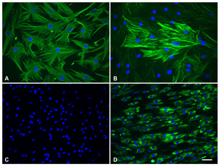
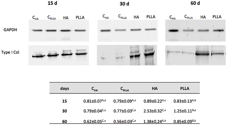
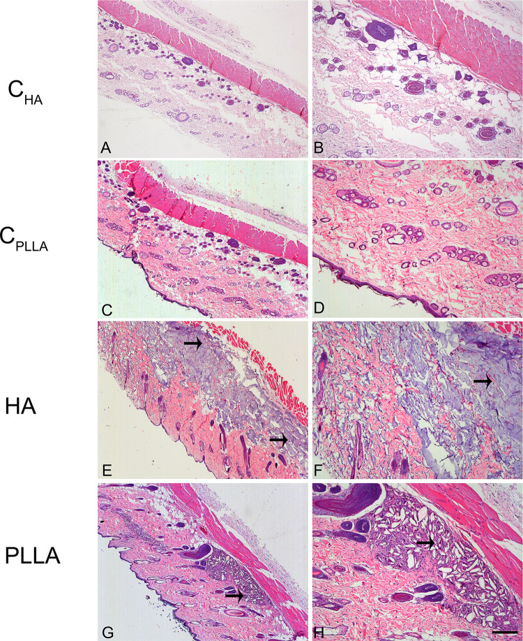
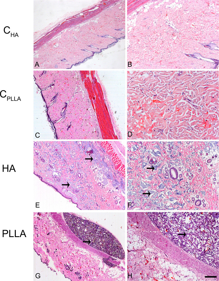
Considering high demand of HA and PLLA as filler material, this study aimed to evaluate their in vitro and in vivo effects. For the in vitro study, human dermal fibroblast cell cultures were supplemented with HA or PLLA for 24, 48, and 72 h. The following parameters were assayed: 1) cell proliferation, 2) cell viability, and 3) quantification of type I collagen by ELISA. For the in vivo study, HA or PLLA was injected in the dermis of Wistar rats and the tissues were collected after 15, 30, and 60 days for histologic evaluation and for quantification of type I collagen by Western blotting. The quantitative data were statistically analyzed using an ANOVA two-way. The significance level was set at 5%.
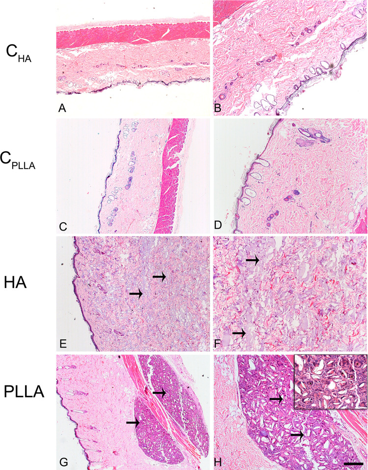
At 72 h, high cell proliferation was observed for HA compared to control (p<0.05). Cultures exposed to PLLA exhibited a reduction in both cell proliferation and viability compared to control in all time points (p<0.05). Type I collagen expression was greater in cultures exposed to HA or PLLA compared to control (p<0.05). Histologic analysis showed the presence of multinucleated cells only in the PLLA group in all experimental time points. Western blotting analysis revealed high content of type I collagen in HA compared to PLLA (p<0.05).
The present study addresses a potentially unfavorable effect of dermal PLLA filler on the fibroblast phenotype, with possible clinical complications, unlike HA.
皮肤老化的特征是表皮和真皮的结构与功能变化,临床上表现为皱纹、弹性丧失和质地粗糙。在此背景下,不同的真皮填充剂已被用于克服与皮肤老化相关的这些负面影响,如透明质酸(HA)和聚左旋乳酸(PLLA)。尽管它们的免疫原性较低,但这些材料在应用后仍可引起炎症反应。
鉴于对HA和PLLA作为填充材料的高需求,本研究旨在评估它们的体外和体内作用。对于体外研究,在人真皮成纤维细胞培养物中添加HA或PLLA 24、48和72小时。检测以下参数:1)细胞增殖,2)细胞活力,3)通过酶联免疫吸附测定法对I型胶原进行定量。对于体内研究,将HA或PLLA注射到Wistar大鼠的真皮中,并在15、30和60天后收集组织进行组织学评估,并通过蛋白质免疫印迹法对I型胶原进行定量。使用双向方差分析对定量数据进行统计分析。显著性水平设定为5%。
在72小时时,与对照组相比,HA组观察到高细胞增殖(p<0.05)。与对照组相比,在所有时间点,暴露于PLLA的培养物在细胞增殖和活力方面均表现出降低(p<0.05)。与对照组相比,暴露于HA或PLLA的培养物中I型胶原表达更高(p<0.05)。组织学分析显示,在所有实验时间点,仅在PLLA组中存在多核细胞。蛋白质免疫印迹分析显示,与PLLA相比,HA中I型胶原含量高(p<0.05)。
本研究揭示了真皮PLLA填充剂对成纤维细胞表型的潜在不利影响,可能存在临床并发症,这与HA不同。