Experimental Imaging Centre, IRCCS San Raffaele Scientific Institute, Via Olgettina, 60, 20132, Milan, Italy.
Medical Physics, IRCCS San Raffaele Scientific Institute, Milan, Italy.
Radiat Oncol. 2020 Oct 17;15(1):240. doi: 10.1186/s13014-020-01684-3.
Methods for the non-invasive quantification of changes in bladder wall thickness as potential predictors of radiation cystitis in pre-clinical research would be desirable. The use of ultrasound for this aim seems promising, but is still relatively unexplored. A method using ultrasound for bladder wall thickness quantification in rats was developed and applied to measure early radiation-induced bladder wall thickness changes.
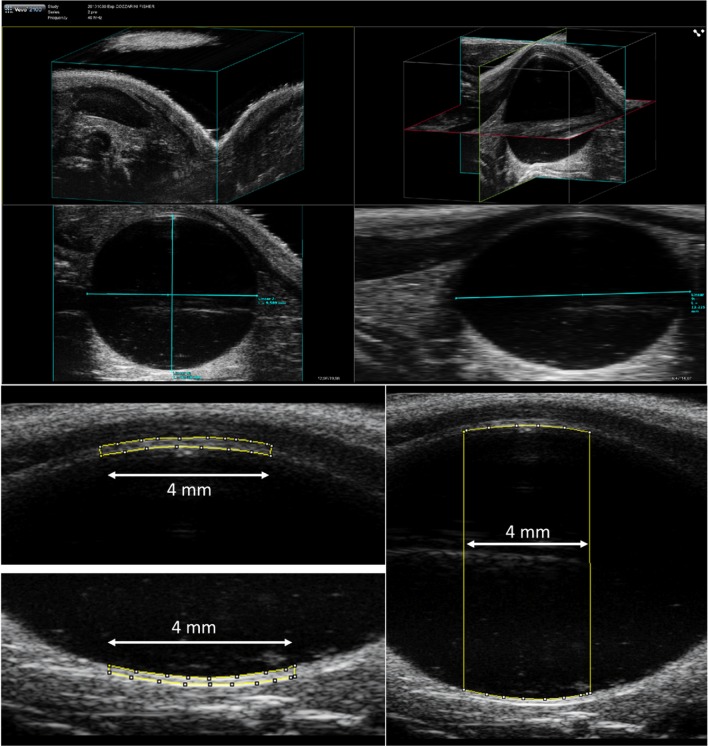
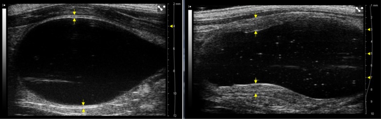
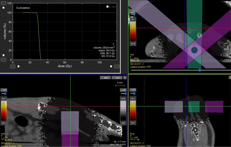
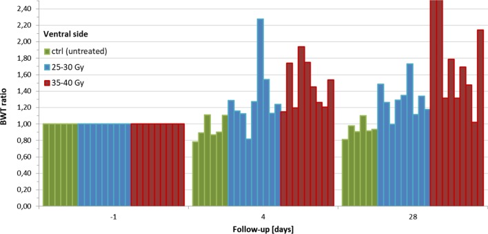
Two groups (n = 9 each) of female Fischer rats were treated with a single radiation dose of 25-30 and 35-40 Gy respectively, using an image-guided micro-irradiator; six untreated rats were monitored as a control group. Empty, half-filled and fully-filled bladder volumes were determined for four non-irradiated rats by measuring axes from ultrasound 3D-images and applying the ellipsoid formula. Mean bladder wall thickness was estimated for both ventral and dorsal bladder sides through the measurement of the bladder wall area along a segment of 4 mm in the central sagittal scan, in order to minimize operator-dependence on the measurement position. Ultrasound acquisitions of all fully-filled rat bladders were also acquired immediately before, and 4 and 28 days after irradiation. Mean bladder wall thickness normalized to the baseline value and corrected for filling were then used to evaluate acute bladder wall thickening and to quantify the dose-effect.
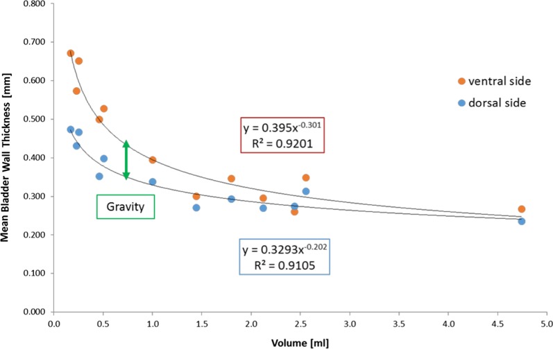
The relationship between mean bladder wall thickness and volume in unirradiated rats showed that for a bladder volume > 1.5 mL the bladder wall thickness is almost constant and equal to 0.30 mm with variations within ± 15%. The average ratios between post and pre irradiation showed a dose-effect relationship. Bladder wall thickening was observed for the 25-30 Gy and 35-40 Gy groups in 2/9 (22%) and 5/9 (56%) cases at day 4 and in 4/9 (44%) and 8/9 (89%) cases at day 28, respectively. The two groups showed significantly different bladder wall thickness both relative to the control group (p < 0.0001) and between them (p = 0.022). The bladder wall thickness increment was on average 1.32 ± 0.41, and was 1.30 ± 0.21 after 25-30 Gy and 1.47 ± 0.29 and 1.90 ± 0.83 after 35-40 Gy at days 4 and 28 respectively.
The feasibility of using ultrasound on a preclinical rat model to detect bladder wall thickness changes after bladder irradiation was demonstrated, and a clear dose-effect relationship was quantified. Although preliminary, these results are promising in addressing the potential role of this non-invasive approach in quantifying radiation cystitis.
在临床前研究中,需要一种非侵入性的方法来定量评估膀胱壁厚度的变化,作为预测放射性膀胱炎的潜在指标。使用超声进行这一目的似乎很有前景,但仍相对未被探索。本研究开发了一种使用超声测量大鼠膀胱壁厚度的方法,并应用于测量早期放射性膀胱壁厚度变化。
两组(每组 9 只)雌性 Fischer 大鼠分别接受单次 25-30 和 35-40 Gy 的放射剂量,使用图像引导的微辐射器;6 只未处理的大鼠作为对照组进行监测。通过测量超声 3D 图像的轴并应用椭圆公式,确定 4 只未照射大鼠的空、半满和全满膀胱容积。通过在中央矢状扫描中测量膀胱壁区域的 4mm 段,估计双侧膀胱壁的平均厚度,以最大程度地减少对测量位置的操作人员依赖性。在照射前、照射后 4 天和 28 天,对所有全充满大鼠膀胱进行超声采集。然后将膀胱壁厚度标准化为基线值,并校正充盈度,以评估急性膀胱壁增厚和量化剂量效应。
在未照射的大鼠中,膀胱壁厚度与体积之间的关系表明,当膀胱体积大于 1.5mL 时,膀胱壁厚度几乎保持不变,等于 0.30mm,且变化幅度在 15%以内。照射后与照射前的平均比值显示出剂量效应关系。25-30Gy 和 35-40Gy 组在第 4 天分别有 2/9(22%)和 5/9(56%)的病例以及在第 28 天分别有 4/9(44%)和 8/9(89%)的病例观察到膀胱壁增厚。两组均与对照组相比(p<0.0001)以及彼此之间(p=0.022)的膀胱壁厚度均有显著差异。膀胱壁厚度增加的平均值为 1.32±0.41,25-30Gy 组为 1.30±0.21,35-40Gy 组分别为 1.47±0.29 和 1.90±0.83,分别在第 4 天和第 28 天。
本研究证明了使用超声在临床前大鼠模型上检测膀胱壁照射后厚度变化的可行性,并定量了明确的剂量效应关系。尽管是初步研究,但这些结果在评估这种非侵入性方法在量化放射性膀胱炎中的潜在作用方面具有很大的潜力。