Department of Molecular and Cellular Physiology, Stanford University, Stanford, CA.
E.L. Ginzton Laboratory, Stanford University, Stanford, CA.
J Gen Physiol. 2020 Nov 2;152(11). doi: 10.1085/jgp.202012672.
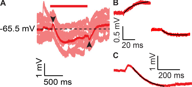
Ultrasound can modulate action potential firing in vivo and in vitro, but the mechanistic basis of this phenomenon is not well understood. To address this problem, we used patch-clamp recording to quantify the effects of focused, high-frequency (43 MHz) ultrasound on evoked action potential firing in CA1 pyramidal neurons in acute rodent hippocampal brain slices. We find that ultrasound can either inhibit or potentiate firing in a spike frequency-dependent manner: at low (near-threshold) input currents and low firing frequencies, ultrasound inhibits firing, while at higher input currents and higher firing frequencies, ultrasound potentiates firing. The net result of these two competing effects is that ultrasound increases the threshold current for action potential firing, the slope of frequency-input curves, and the maximum firing frequency. In addition, ultrasound slightly hyperpolarizes the resting membrane potential, decreases action potential width, and increases the depth of the after-hyperpolarization. All of these results can be explained by the hypothesis that ultrasound activates a sustained potassium conductance. According to this hypothesis, increased outward potassium currents hyperpolarize the resting membrane potential and inhibit firing at near-threshold input currents but potentiate firing in response to higher-input currents by limiting inactivation of voltage-dependent sodium channels during the action potential. This latter effect is a consequence of faster action potential repolarization, which limits inactivation of voltage-dependent sodium channels, and deeper (more negative) after-hyperpolarization, which increases the rate of recovery from inactivation. Based on these results, we propose that ultrasound activates thermosensitive and mechanosensitive two-pore-domain potassium (K2P) channels through heating or mechanical effects of acoustic radiation force. Finite-element modeling of the effects of ultrasound on brain tissue suggests that the effects of ultrasound on firing frequency are caused by a small (<2°C) increase in temperature, with possible additional contributions from mechanical effects.
超声可以调节体内和体外的动作电位发放,但这一现象的机制基础尚不清楚。为了解决这个问题,我们使用膜片钳记录技术来量化聚焦高频(43MHz)超声对急性啮齿动物海马脑片 CA1 锥体神经元诱发动作电位发放的影响。我们发现,超声可以以频率依赖性的方式抑制或增强发放:在低(接近阈)输入电流和低发放频率下,超声抑制发放,而在较高的输入电流和较高的发放频率下,超声增强发放。这两种相互竞争的效应的净结果是,超声增加了动作电位发放的阈电流、频率-输入曲线的斜率和最大发放频率。此外,超声使静息膜电位略微超极化,减小动作电位宽度,并增加后超极化深度。所有这些结果都可以用超声激活持续钾电流的假设来解释。根据这一假设,增加的外向钾电流使静息膜电位超极化,并在接近阈输入电流时抑制发放,但通过限制动作电位期间电压依赖性钠通道的失活,在较高输入电流时增强发放。后一种效应是动作电位更快复极化的结果,这限制了电压依赖性钠通道的失活,并且更深(更负)的后超极化增加了从失活中恢复的速度。基于这些结果,我们提出超声通过热效应或声辐射力的机械效应激活热敏和机械敏感双孔域钾(K2P)通道。超声对脑组织影响的有限元模型表明,超声对发放频率的影响是由小(<2°C)的温度升高引起的,可能还有机械效应的额外贡献。