Department of Thoracic Surgery, 12517Beijing Chao-Yang Hospital, Capital Medical University, Beijing, China.
Technol Cancer Res Treat. 2020 Jan-Dec;19:1533033820957030. doi: 10.1177/1533033820957030.
This study aimed to explore expression profile, its prognostic value, and the potential genomic alterations associated with its dysregulation in lung adenocarcinoma (LUAD) and lung squamous cell carcinoma (LUSC).
Data from The Cancer Genome Atlas (TCGA), The Genotype-Tissue Expression (GTEx), and Kaplan-Meier plotter were used in combination for bioinformatic analysis.
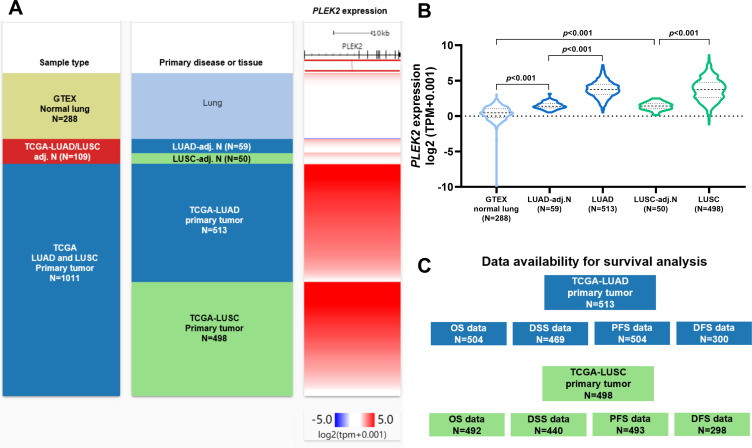
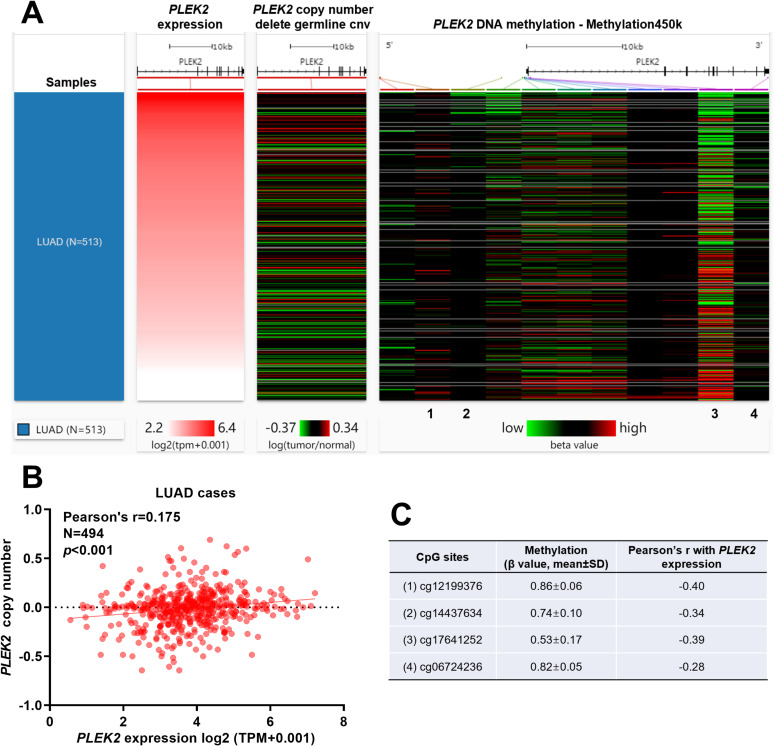
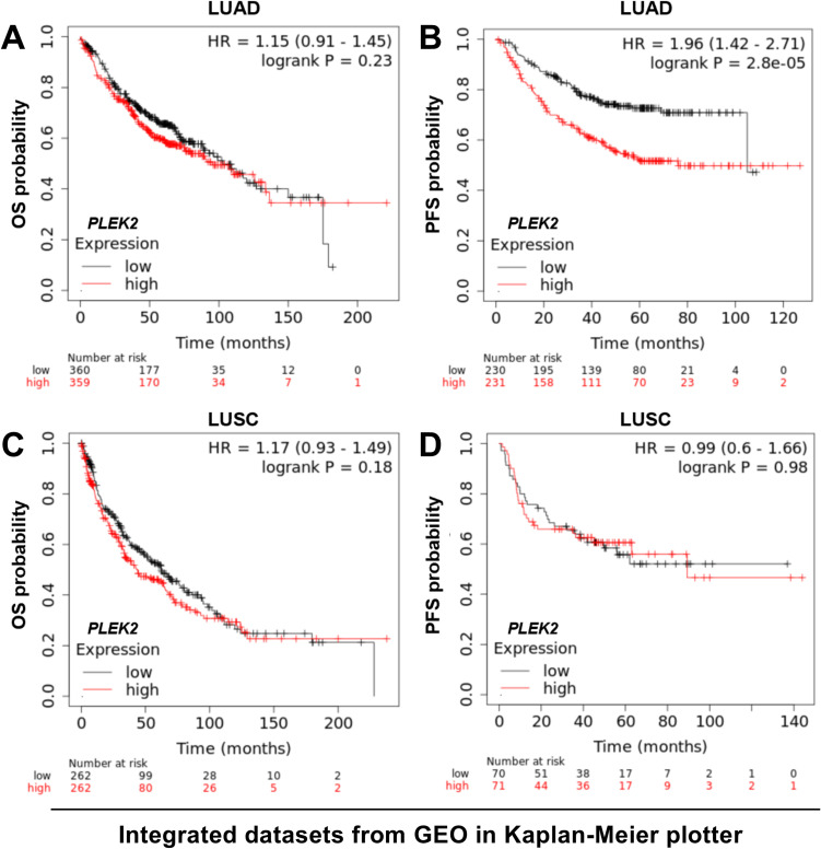
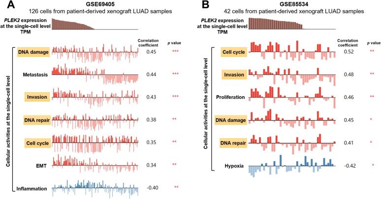
mRNA was significantly upregulated in both LUAD and LUSC compared with their respective normal controls. upregulation showed independent prognostic value in progression-free survival (PFS) (HR: 1.169, 95%CI: 1.033 -1.322, = 0.014). mRNA expression was positively correlated with invasion, cell cycle, DNA damage, and DNA repair of LUAD cells at the single-cell level. Genomic analysis showed that gene-level amplification might not directly lead to increased PLEK2 expression. Methylation profile analysis found 4 CpG sites (cg12199376, cg14437634, cg17641252, and cg06724236) had at least a weakly negative correlation with expression, among which cg12199376, cg14437634 and cg17641252 locate around the first exon of the gene.
Increased expression might be a specific prognostic biomarker of poor PFS in LUAD patients. Its expression had significant positive correlations with invasion, cell cycle, DNA damage, and DNA repair of LUAD cells at the single-cell level. Promoter hypomethylation might be a potential mechanism leading to its upregulation.
本研究旨在探索在肺腺癌(LUAD)和肺鳞状细胞癌(LUSC)中表达谱、其预后价值及其与失调相关的潜在基因组改变。
联合使用癌症基因组图谱(TCGA)、基因型-组织表达(GTEx)和 Kaplan-Meier plotter 进行生物信息学分析。
与相应的正常对照相比,mRNA 在 LUAD 和 LUSC 中均显著上调。上调在无进展生存期(PFS)中具有独立的预后价值(HR:1.169,95%CI:1.033-1.322,P=0.014)。mRNA 表达与 LUAD 细胞在单细胞水平的侵袭、细胞周期、DNA 损伤和 DNA 修复呈正相关。基因组分析表明,基因水平扩增可能不会直接导致 PLEK2 表达增加。甲基化谱分析发现 4 个 CpG 位点(cg12199376、cg14437634、cg17641252 和 cg06724236)与基因表达至少存在弱负相关,其中 cg12199376、cg14437634 和 cg17641252 位于基因的第一个外显子周围。
表达增加可能是 LUAD 患者 PFS 不良的特异性预后生物标志物。其表达与 LUAD 细胞在单细胞水平的侵袭、细胞周期、DNA 损伤和 DNA 修复呈显著正相关。启动子低甲基化可能是导致其上调的潜在机制。