Millstead Luke, Jayakody Hiranya, Patel Harsh, Kaura Vihaan, Petrie Paul R, Tomasetig Florence, Whitty Mark
School of Mechanical and Manufacturing Engineering, University of New South Wales, Sydney, NSW, Australia.
Crop Sciences Division, South Australian Research and Development Institute, Waite Campus, Urrbrae, SA, Australia.
Front Plant Sci. 2020 Sep 25;11:580389. doi: 10.3389/fpls.2020.580389. eCollection 2020.
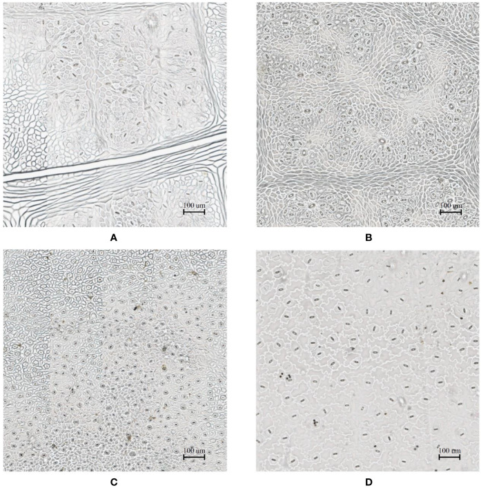
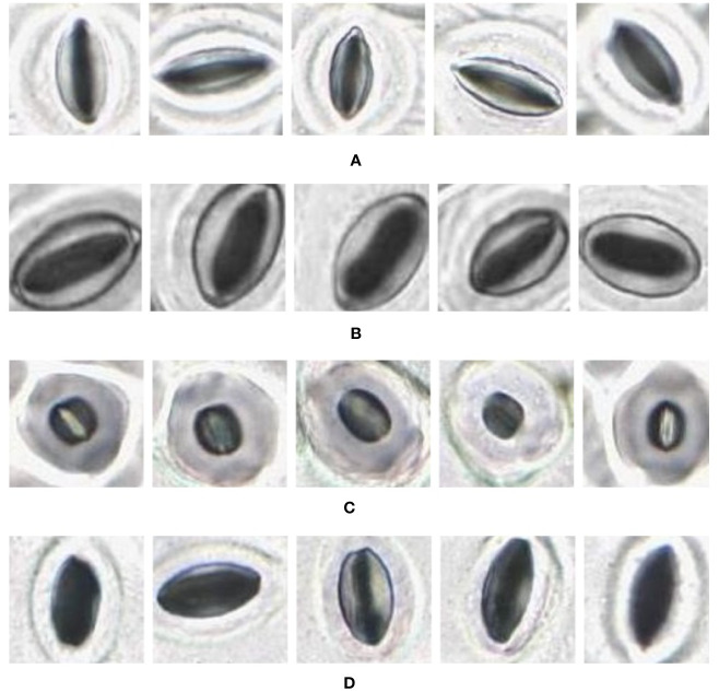
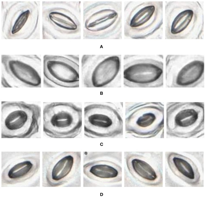
Digital image processing is commonly used in plant health and growth analysis, aiming to improve research efficiency and repeatability. One focus is analysing the morphology of stomata, with the aim to better understand the regulation of gas exchange, its link to photosynthesis and water use and how they are influenced by climatic conditions. Despite the key role played by these cells, their microscopic analysis is largely manual, requiring intricate sample collection, laborious microscope application and the manual operation of a graphical user interface to identify and measure stomata. This research proposes a simple, end-to-end solution which enables automatic analysis of stomata by introducing key changes to imaging techniques, stomata detection as well as stomatal pore area calculation. An optimal procedure was developed for sample collection and imaging by investigating the suitability of using an automatic microscope slide scanner to image nail polish imprints. The use of the slide scanner allows the rapid collection of high-quality images from entire samples with minimal manual effort. A convolutional neural network was used to automatically detect stomata in the input image, achieving average precision, recall and F-score values of 0.79, 0.85, and 0.82 across four plant species. A novel binary segmentation and stomatal cross section analysis method is developed to estimate the pore boundary and calculate the associated area. The pore estimation algorithm correctly identifies stomata pores 73.72% of the time. Ultimately, this research presents a fast and simplified method of stomatal assay generation requiring minimal human intervention, enhancing the speed of acquiring plant health information.
数字图像处理常用于植物健康与生长分析,旨在提高研究效率和可重复性。一个重点是分析气孔形态,以便更好地理解气体交换的调节、其与光合作用和水分利用的联系,以及它们如何受到气候条件的影响。尽管这些细胞发挥着关键作用,但对它们的微观分析在很大程度上是手动的,需要复杂的样本采集、费力地使用显微镜以及通过图形用户界面进行手动操作来识别和测量气孔。本研究提出了一种简单的端到端解决方案,通过对成像技术、气孔检测以及气孔孔径计算引入关键变化,实现气孔的自动分析。通过研究使用自动显微镜载玻片扫描仪对指甲油印记进行成像的适用性,开发了一种用于样本采集和成像的最佳程序。使用载玻片扫描仪可以用最少的人工操作从整个样本中快速采集高质量图像。利用卷积神经网络自动检测输入图像中的气孔,在四种植物物种上实现的平均精度、召回率和F值分别为0.79、0.85和0.82。开发了一种新颖的二值分割和气孔横截面分析方法来估计气孔边界并计算相关面积。气孔估计算法正确识别气孔的概率为73.72%。最终,本研究提出了一种快速且简化的气孔分析方法,所需人工干预最少,提高了获取植物健康信息的速度。