• 文献检索
  • 文档翻译
  • 深度研究
  • 学术资讯
  • Suppr Zotero 插件Zotero 插件
  • 邀请有礼
  • 套餐&价格
  • 历史记录
应用&插件
Suppr Zotero 插件Zotero 插件浏览器插件Mac 客户端Windows 客户端微信小程序
定价
高级版会员购买积分包购买API积分包
服务
文献检索文档翻译深度研究API 文档MCP 服务
关于我们
关于 Suppr公司介绍联系我们用户协议隐私条款
关注我们

Suppr 超能文献

核心技术专利:CN118964589B侵权必究
粤ICP备2023148730 号-1Suppr @ 2026

文献检索

告别复杂PubMed语法,用中文像聊天一样搜索,搜遍4000万医学文献。AI智能推荐,让科研检索更轻松。

立即免费搜索

文件翻译

保留排版,准确专业,支持PDF/Word/PPT等文件格式,支持 12+语言互译。

免费翻译文档

深度研究

AI帮你快速写综述,25分钟生成高质量综述,智能提取关键信息,辅助科研写作。

立即免费体验

踝关节和足部的磁共振成像。

Magnetic resonance imaging of the ankle and foot.

作者信息

Gorbachova Tetyana

机构信息

Einstein Medical Center, USA.

出版信息

Pol J Radiol. 2020 Sep 18;85:e532-e549. doi: 10.5114/pjr.2020.99472. eCollection 2020.

DOI:10.5114/pjr.2020.99472
PMID:33101556
原文链接:https://pmc.ncbi.nlm.nih.gov/articles/PMC7571512/
Abstract

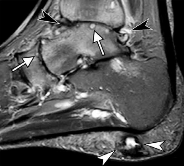
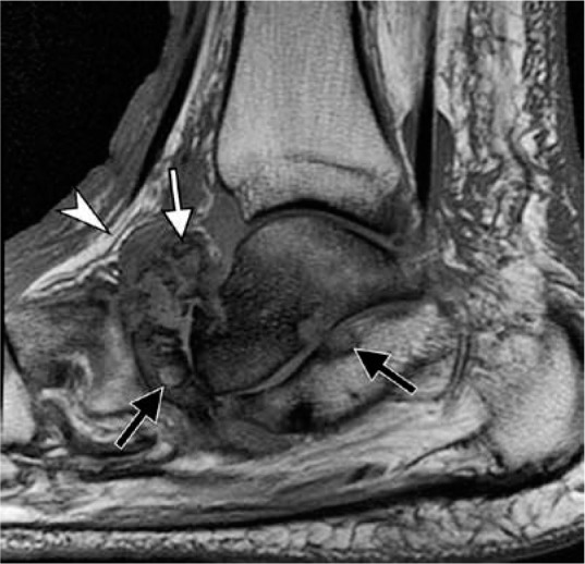
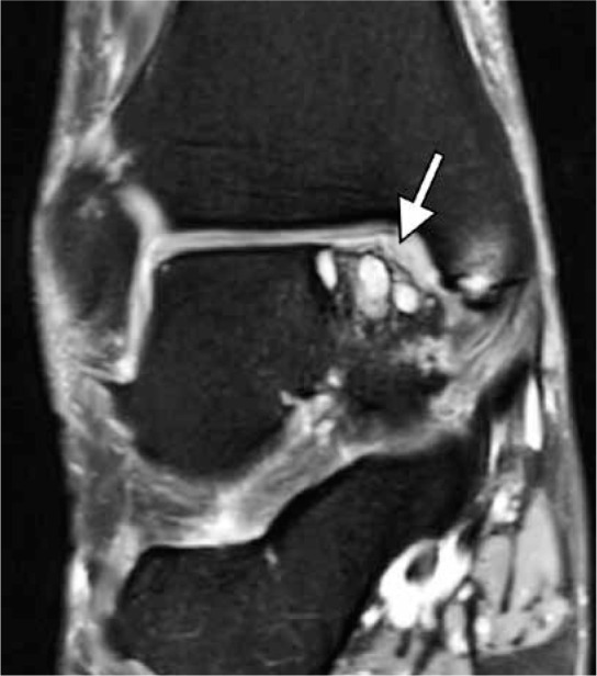
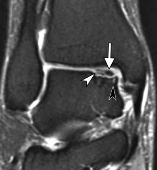
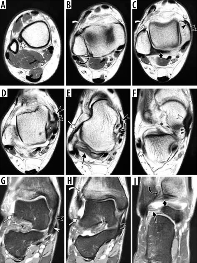
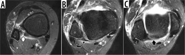
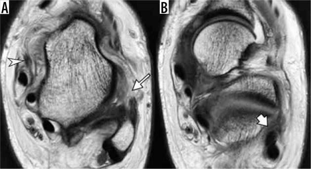
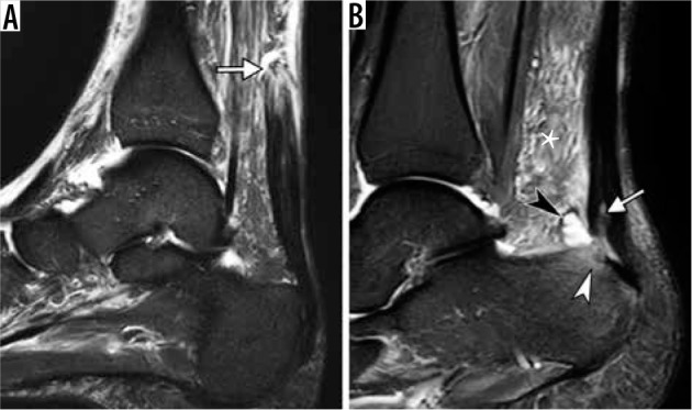
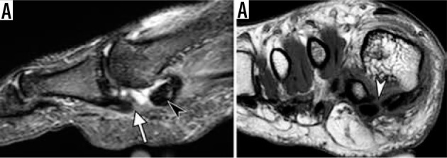
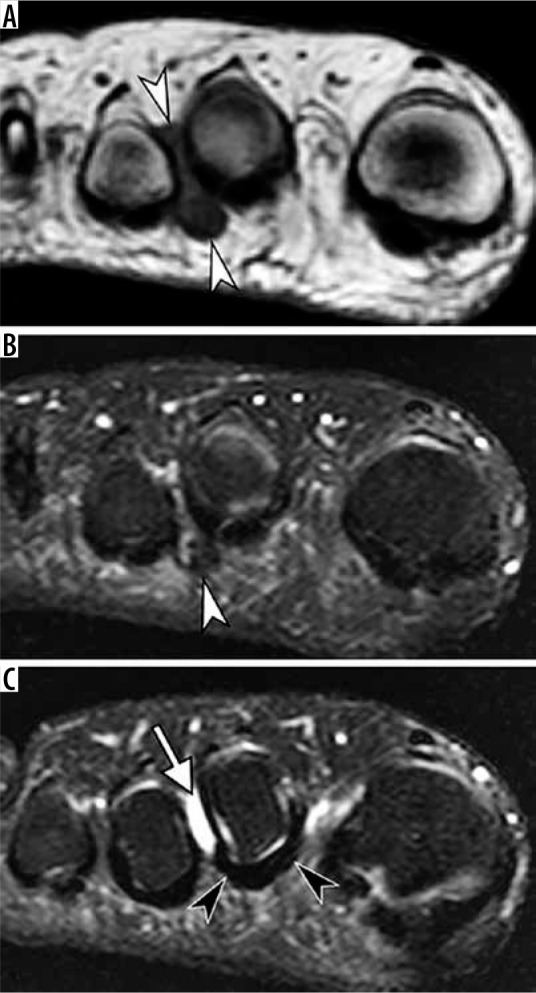
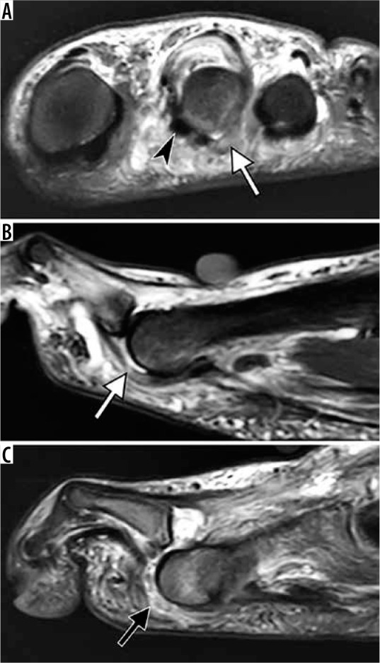
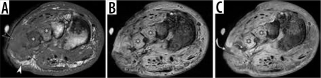
This article reviews the magnetic resonance imaging (MRI) findings of the normal anatomy and various pathologic conditions of the ankle and foot commonly encountered in clinical practice. The spectrum of entities discussed includes osseous and osteochondral injuries, ligamentous injuries, common traumatic and degenerative tendon pathology, abnormalities of transverse tarsal joint (Chopart) and tarsometatarsal joint (Lisfranc) complexes, pathological conditions affecting capsuloligamentous structures of the great toe and lesser toes, as well as pedal infection, with a focus on diabetic osteomyelitis and neuropathic osteoarthropathy.

摘要

本文综述了临床实践中常见的踝关节和足部正常解剖结构及各种病理状况的磁共振成像(MRI)表现。所讨论的病变范围包括骨与骨软骨损伤、韧带损伤、常见的创伤性和退行性肌腱病变、跗横关节(Chopart关节)和跗跖关节(Lisfranc关节)复合体异常、影响拇趾和小趾关节囊韧带结构的病理状况以及足部感染,重点关注糖尿病性骨髓炎和神经性骨关节病。

https://cdn.ncbi.nlm.nih.gov/pmc/blobs/5a12/7571512/b5fd6a8299c5/PJR-85-41908-g028.jpg
https://cdn.ncbi.nlm.nih.gov/pmc/blobs/5a12/7571512/3d3bcf9a578d/PJR-85-41908-g003.jpg
https://cdn.ncbi.nlm.nih.gov/pmc/blobs/5a12/7571512/257e8bd129e9/PJR-85-41908-g004.jpg
https://cdn.ncbi.nlm.nih.gov/pmc/blobs/5a12/7571512/ffb05870716f/PJR-85-41908-g005.jpg
https://cdn.ncbi.nlm.nih.gov/pmc/blobs/5a12/7571512/6f3d056e6fc1/PJR-85-41908-g006.jpg
https://cdn.ncbi.nlm.nih.gov/pmc/blobs/5a12/7571512/e3ea70d5493b/PJR-85-41908-g007.jpg
https://cdn.ncbi.nlm.nih.gov/pmc/blobs/5a12/7571512/052dd9e406c6/PJR-85-41908-g008.jpg
https://cdn.ncbi.nlm.nih.gov/pmc/blobs/5a12/7571512/8a33c02798ca/PJR-85-41908-g009.jpg
https://cdn.ncbi.nlm.nih.gov/pmc/blobs/5a12/7571512/6bf6ca593f36/PJR-85-41908-g010.jpg
https://cdn.ncbi.nlm.nih.gov/pmc/blobs/5a12/7571512/1f6aa0a54dfa/PJR-85-41908-g011.jpg
https://cdn.ncbi.nlm.nih.gov/pmc/blobs/5a12/7571512/96227c4a13f1/PJR-85-41908-g012.jpg
https://cdn.ncbi.nlm.nih.gov/pmc/blobs/5a12/7571512/74c21394dad9/PJR-85-41908-g013.jpg
https://cdn.ncbi.nlm.nih.gov/pmc/blobs/5a12/7571512/9823e51e1fb7/PJR-85-41908-g014.jpg
https://cdn.ncbi.nlm.nih.gov/pmc/blobs/5a12/7571512/3ff28c23e49b/PJR-85-41908-g015.jpg
https://cdn.ncbi.nlm.nih.gov/pmc/blobs/5a12/7571512/611a3a3f183a/PJR-85-41908-g016.jpg
https://cdn.ncbi.nlm.nih.gov/pmc/blobs/5a12/7571512/2b6912ae72f7/PJR-85-41908-g017.jpg
https://cdn.ncbi.nlm.nih.gov/pmc/blobs/5a12/7571512/efffde5af3d4/PJR-85-41908-g018.jpg
https://cdn.ncbi.nlm.nih.gov/pmc/blobs/5a12/7571512/3f5df19c7984/PJR-85-41908-g019.jpg
https://cdn.ncbi.nlm.nih.gov/pmc/blobs/5a12/7571512/2ad1d67a869b/PJR-85-41908-g020.jpg
https://cdn.ncbi.nlm.nih.gov/pmc/blobs/5a12/7571512/818d7ff975bc/PJR-85-41908-g021.jpg
https://cdn.ncbi.nlm.nih.gov/pmc/blobs/5a12/7571512/c8047fe38c0f/PJR-85-41908-g022.jpg
https://cdn.ncbi.nlm.nih.gov/pmc/blobs/5a12/7571512/48fdbdf89603/PJR-85-41908-g023.jpg
https://cdn.ncbi.nlm.nih.gov/pmc/blobs/5a12/7571512/3dc4b24c669a/PJR-85-41908-g024.jpg
https://cdn.ncbi.nlm.nih.gov/pmc/blobs/5a12/7571512/8d8022f4718f/PJR-85-41908-g025.jpg
https://cdn.ncbi.nlm.nih.gov/pmc/blobs/5a12/7571512/2384b60b5079/PJR-85-41908-g026.jpg
https://cdn.ncbi.nlm.nih.gov/pmc/blobs/5a12/7571512/03f7cd485d0e/PJR-85-41908-g027.jpg
https://cdn.ncbi.nlm.nih.gov/pmc/blobs/5a12/7571512/b5fd6a8299c5/PJR-85-41908-g028.jpg

相似文献

1
Magnetic resonance imaging of the ankle and foot.踝关节和足部的磁共振成像。
Pol J Radiol. 2020 Sep 18;85:e532-e549. doi: 10.5114/pjr.2020.99472. eCollection 2020.
2
MR Imaging of the Midfoot Including Chopart and Lisfranc Joint Complexes.包括Chopart和Lisfranc关节复合体在内的中足的磁共振成像
Magn Reson Imaging Clin N Am. 2017 Feb;25(1):95-125. doi: 10.1016/j.mric.2016.08.006.
3
Imaging in Foot and Ankle Instability.足踝不稳的影像学检查
Foot Ankle Clin. 2018 Dec;23(4):499-522.e28. doi: 10.1016/j.fcl.2018.07.011.
4
JOURNAL CLUB: MRI Evaluation of Midtarsal (Chopart) Sprain in the Setting of Acute Ankle Injury.期刊俱乐部:急性踝关节损伤中跗中(Chopart)扭伤的 MRI 评估。
AJR Am J Roentgenol. 2018 Feb;210(2):386-395. doi: 10.2214/AJR.17.18503. Epub 2017 Nov 7.
5
CT and MR Imaging of the Postoperative Ankle and Foot.术后踝关节和足部的CT与磁共振成像
Radiographics. 2016 Oct;36(6):1828-1848. doi: 10.1148/rg.2016160016.
6
Imaging evaluation of traumatic ligamentous injuries of the ankle and foot.踝与足部创伤性韧带损伤的影像学评估
Radiol Clin North Am. 2013 May;51(3):455-78. doi: 10.1016/j.rcl.2012.11.004. Epub 2013 Mar 25.
7
Normal Anatomy and Traumatic Injury of the Midtarsal (Chopart) Joint Complex: An Imaging Primer.中跗(Chopart)关节复合体的正常解剖结构和外伤性损伤:影像学基础。
Radiographics. 2019 Jan-Feb;39(1):136-152. doi: 10.1148/rg.2019180102. Epub 2018 Nov 30.
8
[Injuries of ligaments and tendons of foot and ankle : What every radiologist should know].[足踝部韧带和肌腱损伤:每位放射科医生都应了解的内容]
Radiologe. 2018 May;58(5):415-421. doi: 10.1007/s00117-018-0383-7.
9
Imaging of Chopart (Midtarsal) Joint Complex: Normal Anatomy and Posttraumatic Findings.Chopart(跗中)关节复合体的影像学表现:正常解剖与创伤后表现。
AJR Am J Roentgenol. 2018 Aug;211(2):416-425. doi: 10.2214/AJR.17.19310. Epub 2018 Jun 21.
10
Magnetic resonance imaging of the foot and ankle: correlation of normal anatomy with pathologic conditions.
Foot Ankle. 1991 Apr;11(5):289-305. doi: 10.1177/107110079101100506.

引用本文的文献

1
Association between clinical and MRI-detected imaging findings for people with midfoot pain, a cross-sectional study.中足疼痛患者临床与MRI检测影像学表现之间的关联:一项横断面研究
J Foot Ankle Res. 2025 Mar;18(1):e70019. doi: 10.1002/jfa2.70019.
2
MRI Evaluation of Traumatic and Non-traumatic Pathologies of the Ankle Joint and Hindfoot: A Single-Center Observational Study.踝关节和后足创伤性与非创伤性病变的MRI评估:一项单中心观察性研究
Cureus. 2024 Aug 18;16(8):e67103. doi: 10.7759/cureus.67103. eCollection 2024 Aug.
3
Correlation between Ankle Imaging Findings and Self-Reported Outcomes: A Longitudinal Assessment in Patients with Tibiofibular Diastasis.

本文引用的文献

1
Predicting osteomyelitis in patients whose initial MRI demonstrated bone marrow edema without corresponding T1 signal marrow replacement.预测初始 MRI 显示骨髓水肿但无相应 T1 信号骨髓置换的患者发生骨髓炎。
Skeletal Radiol. 2020 Aug;49(8):1239-1247. doi: 10.1007/s00256-020-03396-x. Epub 2020 Mar 4.
2
Adult Acquired Flatfoot Deformity: Anatomy, Biomechanics, Staging, and Imaging Findings.成人获得性平足畸形:解剖、生物力学、分期和影像学表现。
Radiographics. 2019 Sep-Oct;39(5):1437-1460. doi: 10.1148/rg.2019190046.
3
Nomenclature of Subchondral Nonneoplastic Bone Lesions.
踝关节影像学表现与自我报告结果之间的相关性:胫腓骨分离患者的纵向评估
J Clin Med. 2023 Nov 22;12(23):7239. doi: 10.3390/jcm12237239.
4
Talus bone: normal anatomy, anatomical variations and clinical correlations.距骨:正常解剖、解剖变异与临床关联。
Anat Sci Int. 2023 Jul;98(3):391-406. doi: 10.1007/s12565-023-00712-y. Epub 2023 Apr 5.
5
Imaging of osteoarthritis from the ankle through the midfoot.从踝关节到中足部的骨关节炎影像学表现。
Skeletal Radiol. 2023 Nov;52(11):2239-2257. doi: 10.1007/s00256-023-04287-7. Epub 2023 Feb 4.
6
Isolated, periosteal stripping injuries of the Flexor Retinaculum: Case series of 3 patients with clinico-radiological review.屈肌支持带孤立性骨膜剥离损伤:3例患者临床与影像学回顾的病例系列
J Clin Orthop Trauma. 2022 Nov 24;36:102082. doi: 10.1016/j.jcot.2022.102082. eCollection 2023 Jan.
7
Magnetic Resonance Imaging of Ankle Disorders in Adult Nigerians in Lagos.拉各斯成年尼日利亚人的踝关节疾病的磁共振成像
J West Afr Coll Surg. 2022 Apr-Jun;12(2):81-87. doi: 10.4103/jwas.jwas_134_22. Epub 2022 Aug 27.
8
Magnetic resonance imaging of musculoskeletal infections.肌肉骨骼感染的磁共振成像
Pol J Radiol. 2022 Mar 5;87:e141-e162. doi: 10.5114/pjr.2022.113825. eCollection 2022.
9
Imaging and Treatment of Posttraumatic Ankle and Hindfoot Osteoarthritis.创伤后踝与后足骨关节炎的影像学检查与治疗
J Clin Med. 2021 Dec 13;10(24):5848. doi: 10.3390/jcm10245848.
骨软骨下非肿瘤性骨病变命名。
AJR Am J Roentgenol. 2019 Nov;213(5):963-982. doi: 10.2214/AJR.19.21571. Epub 2019 Jul 24.
4
The peroneus longus muscle and tendon: a review of its anatomy and pathology.腓骨长肌及其肌腱:解剖与病理研究综述。
Skeletal Radiol. 2019 Sep;48(9):1329-1344. doi: 10.1007/s00256-019-3168-9. Epub 2019 Feb 15.
5
Normal Anatomy and Traumatic Injury of the Midtarsal (Chopart) Joint Complex: An Imaging Primer.中跗(Chopart)关节复合体的正常解剖结构和外伤性损伤:影像学基础。
Radiographics. 2019 Jan-Feb;39(1):136-152. doi: 10.1148/rg.2019180102. Epub 2018 Nov 30.
6
Insertional anatomy of peroneal brevis and longus tendon - A cadaveric study.比目鱼肌短肌和长肌肌腱的插入解剖结构——尸体研究。
Foot Ankle Surg. 2019 Oct;25(5):636-639. doi: 10.1016/j.fas.2018.07.005. Epub 2018 Jul 23.
7
Not One of the Usual Suspects: MRI Features of Insertional Lateral Cord Plantar Fasciopathy.非常见的致病因素之一:MRI 对插入性外侧跟骨足底筋膜炎的特征表现。
AJR Am J Roentgenol. 2018 Nov;211(5):W252-W256. doi: 10.2214/AJR.17.19218. Epub 2018 Sep 21.
8
Added value of MRI to X-ray in guiding the extent of surgical resection in diabetic forefoot osteomyelitis: a review of pathologically proven, surgically treated cases.MRI对X线在指导糖尿病足前部骨髓炎手术切除范围方面的附加价值:对经病理证实并接受手术治疗病例的综述
Skeletal Radiol. 2019 Mar;48(3):405-411. doi: 10.1007/s00256-018-3045-y. Epub 2018 Aug 22.
9
Osteochondral Lesions of the Knee: Differentiating the Most Common Entities at MRI.膝关节骨软骨病变:MRI 鉴别最常见病变。
Radiographics. 2018 Sep-Oct;38(5):1478-1495. doi: 10.1148/rg.2018180044. Epub 2018 Aug 17.
10
Acute Fracture of the Anterior Process of Calcaneus: Does It Herald a More Advanced Injury to Chopart Joint?跟骨前突急性骨折:是否预示着 Chopart 关节有更严重的损伤?
AJR Am J Roentgenol. 2018 May;210(5):1123-1130. doi: 10.2214/AJR.17.18678. Epub 2018 Mar 23.