Department of Developmental Genetics, Institute of Advanced Medicine, Wakayama Medical University, Kimiidera 811-1, Wakayama City, Wakayama, 641-8509, Japan.
Laboratory of Hygienic Chemistry and Molecular Toxicology, Gifu Pharmaceutical University, 1-25-4 Daigaku-nishi, Gifu-City, Gifu, 501-1196, Japan.
Sci Rep. 2020 Oct 26;10(1):18251. doi: 10.1038/s41598-020-75184-5.
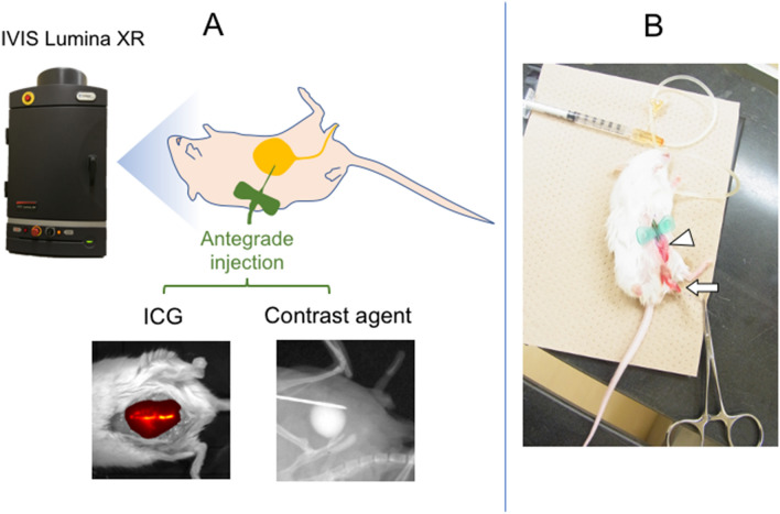
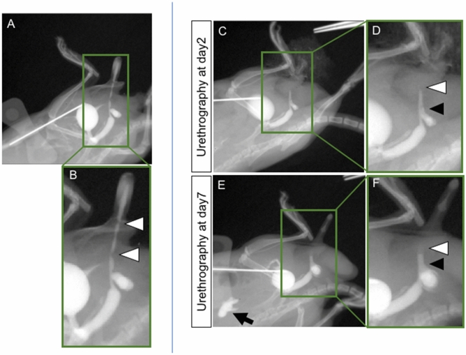
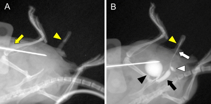
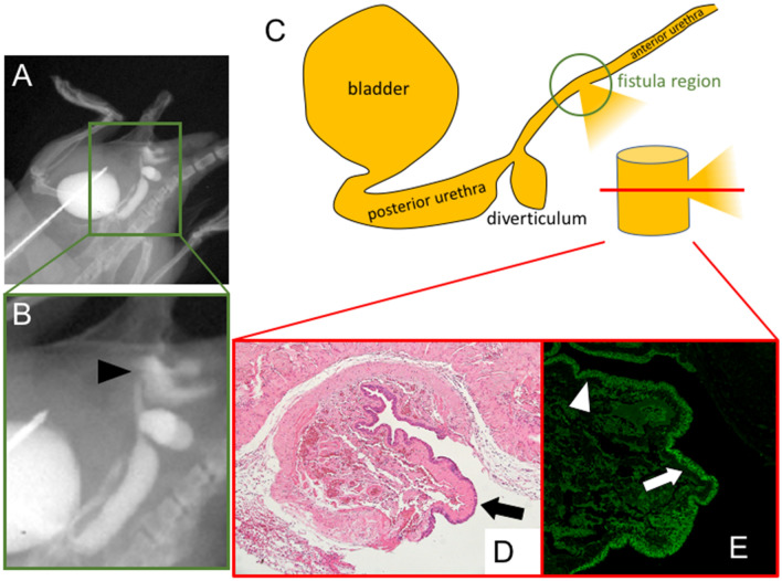
Visualization of the surgically operated tissues is vital to improve surgical model animals including mouse. Urological surgeries for urethra include series of fine manipulations to treat the increasing number of birth defects such as hypospadias. Hence visualization of the urethral status is vital. Inappropriate urethral surgical procedure often leads to the incomplete wound healing and subsequent formation of urethro-cutaneous fistula or urethral stricture. Application of indocyanine green mediated visualization of the urethra was first performed in the current study. Indocyanine green revealed the bladder but not the urethral status in mouse. Antegrade injection of contrast agent into the bladder enabled to detect the urethral status in vivo. The visualization of the leakage of contrast agent from the operated region was shown as the state of urethral fistula in the current hypospadias mouse model and urethral stricture was also revealed. A second trial for contrast agent was performed after the initial operation and a tendency of accelerated urethral stricture was observed. Thus, assessment of post-surgical conditions of urogenital tissues can be improved by the current analyses on the urethral status.
手术组织的可视化对于改进包括小鼠在内的手术模型动物至关重要。尿道的泌尿科手术包括一系列精细的操作,以治疗日益增多的出生缺陷,如尿道下裂。因此,尿道状况的可视化至关重要。不适当的尿道手术程序通常会导致伤口愈合不完全,并随后形成尿道皮瘘或尿道狭窄。本研究首次应用吲哚菁绿介导的尿道可视化。吲哚菁绿显示了膀胱,但没有显示小鼠的尿道状况。将造影剂顺行注射到膀胱中,可以在体内检测到尿道状况。从手术区域漏出的造影剂的可视化显示为当前尿道下裂小鼠模型中的尿道瘘状态,并且还显示了尿道狭窄。在初次手术后进行了第二次造影剂试验,观察到尿道狭窄加速的趋势。因此,通过当前对尿道状况的分析,可以改善泌尿组织的术后状况评估。