Department of Pediatric Surgery, University Medical Center Hamburg-Eppendorf, UKE Medical School, Martinistrasse 52, 20246, Hamburg, Germany.
Sci Rep. 2020 Oct 26;10(1):18240. doi: 10.1038/s41598-020-74370-9.
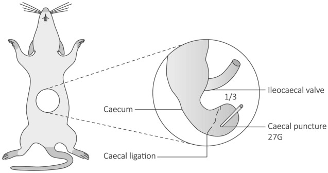
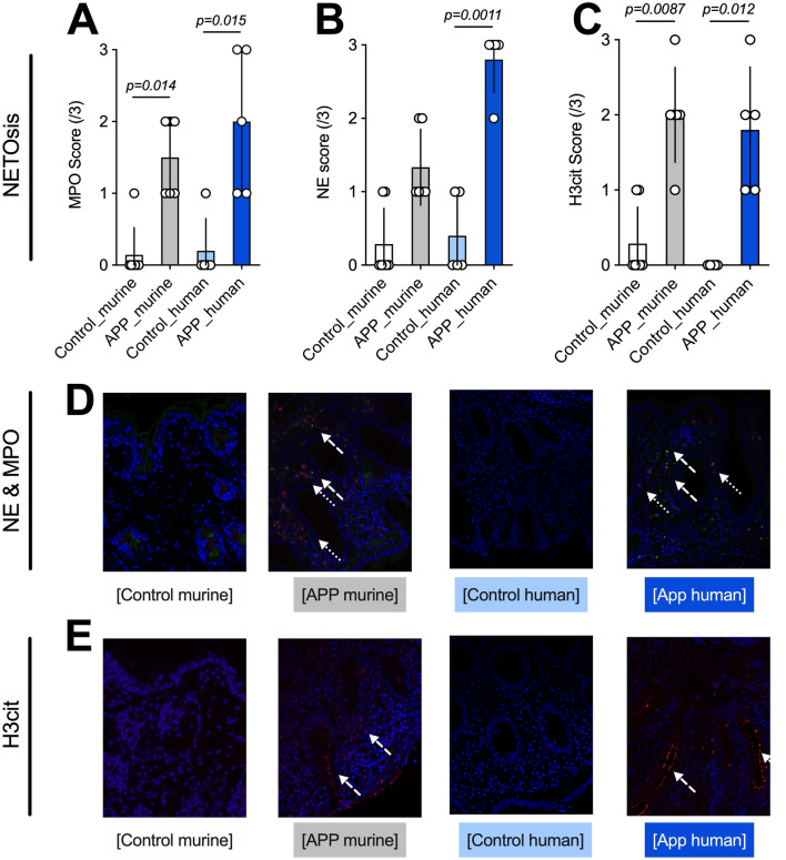
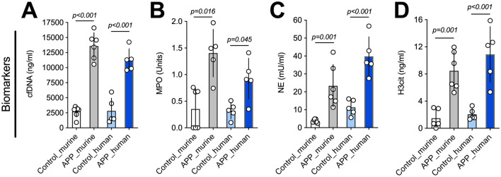
Appendicitis is one of the most frequent emergencies in pediatric surgery, yet current biomarkers for diagnosis are unspecific and have low predictive values. As neutrophils and extracellular traps (ETs) are an essential component of the immune defense against bacterial infections, and appendicitis is considered an inflammation reaction of the appendix, we hypothesized that neutrophil activation and NET formation play an essential role in appendicitis development and maintenance. Therefore, this pilot study aimed to establish a murine model of appendicitis and to evaluate ETs markers to diagnose appendicitis in mice and humans. The study used 20 (12 appendicitis- and 8 controls) 6-week old mice which underwent advanced appendicitis induction using a modified caecal ligation puncture procedure. During the study, cell-free DNA, neutrophil elastase (NE), myeloperoxidase (MPO), and citrullinated Histone H3 (H3cit) were assessed. Additionally, samples of 5 children with histologically confirmed appendicitis and 5 matched controls with catarrhal appendicitis, were examined for the same biomarkers. Moreover, NE, MPO, and H3cit were assessed histologically via immunofluorescence in mice and humans. All mice in the appendicitis group developed an advanced form of appendicitis with focal peritonitis. In mice and humans with appendicitis, markers of neutrophil activation and ETs formation (especially cfDNA, NE and H3cit) were significantly elevated in blood and tissue compared to controls. Ultimately, biomarkers correlated extremely well with tissue expression and thus disease severity. It appears that neutrophil activation and possibly NETs contribute to appendicitis development and biomarkers of neutrophil activation and ET formation reflect disease severity and thus could be used as biomarkers for appendicitis. However, large prospective clinical studies are needed to confirm our findings.
阑尾炎是小儿外科最常见的急症之一,但目前用于诊断的生物标志物特异性不强,预测价值低。由于中性粒细胞和细胞外陷阱 (NETs) 是抵御细菌感染的免疫防御的重要组成部分,而阑尾炎被认为是阑尾的炎症反应,我们假设中性粒细胞的激活和 NET 的形成在阑尾炎的发展和维持中起着重要作用。因此,本研究旨在建立一种阑尾炎的小鼠模型,并评估 ETs 标志物以诊断小鼠和人类的阑尾炎。本研究使用了 20 只(12 只阑尾炎和 8 只对照)6 周龄的小鼠,通过改良的盲肠结扎穿刺术诱导阑尾炎。在研究过程中,评估了无细胞 DNA、中性粒细胞弹性蛋白酶 (NE)、髓过氧化物酶 (MPO) 和瓜氨酸化组蛋白 H3 (H3cit)。此外,还检查了 5 例经组织学证实的阑尾炎患儿和 5 例伴有卡他性阑尾炎的匹配对照者的相同生物标志物。此外,通过免疫荧光法在小鼠和人类中评估了 NE、MPO 和 H3cit 的组织学表现。阑尾炎组的所有小鼠均发展为具有局灶性腹膜炎的晚期阑尾炎。在患有阑尾炎的小鼠和人类中,与对照组相比,血液和组织中的中性粒细胞激活和 NET 形成标志物(尤其是 cfDNA、NE 和 H3cit)显著升高。最终,生物标志物与组织表达高度相关,因此与疾病严重程度相关。似乎中性粒细胞的激活和可能的 NET 有助于阑尾炎的发展,并且中性粒细胞激活和 NET 形成的生物标志物反映了疾病的严重程度,因此可以作为阑尾炎的生物标志物。然而,需要进行大规模的前瞻性临床研究来证实我们的发现。