Department of Ophthalmology, IRCCS-Fondazione Bietti, Rome, Italy.
Unit of Endocrinology, Diabetes and Metabolism, S. Giovanni Calibita Fatebenefratelli Hospital, Department of Systems Medicine, University of Rome Tor Vergata, Rome, Italy.
Sci Rep. 2020 Oct 26;10(1):18266. doi: 10.1038/s41598-020-75416-8.
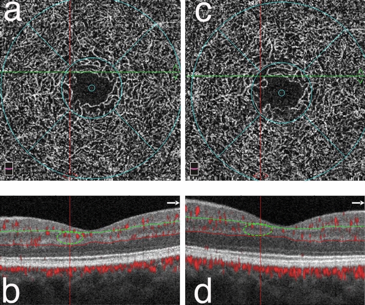
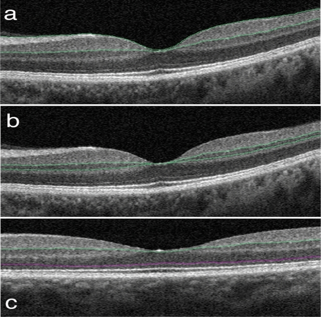
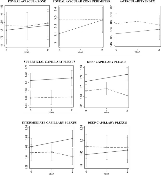
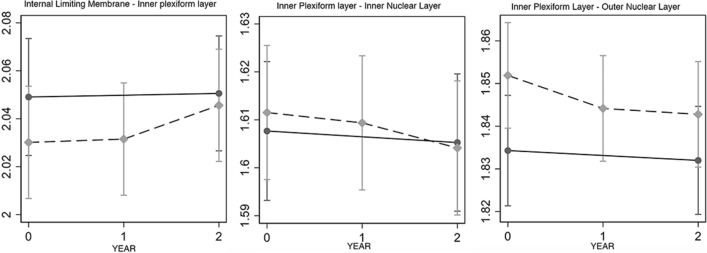
This study aimed to explore differences in vascular and structural parameters using optical coherence tomography angiography in patients with type 1 diabetes (DM1) with mild signs of diabetic retinopathy (DR) over a two-year follow-up period. Parafoveal vessel density (PVD) and foveal avascular zone (FAZ) area were analyzed. The thickness of three predefined retinal slabs was measured, including the inner limiting membrane (ILM)-inner plexiform layer (IPL), IPL-inner nuclear layer (INL), and the IPL-outer nuclear layer (ONL). Twenty-two patients with DM1 and 21 controls were included. There was no significant difference in the FAZ area, perimeter and acircularity index between cohorts over time. Baseline superficial capillary plexus PVD was approximately 10% lower in patients with diabetes than in controls (p = 0.001), and was 12% lower at 2 years (p = 0.002). There was no difference in the annual linear trend between the groups (- 0.5% in diabetics vs. controls, p = 0.736). Baseline deep capillary plexus (DCP) PVD was slightly lower in diabetics than in controls (- 4.4%, p = 0.047) and the difference increased at 2 years (- 12.6%, p < 0.001). The annual linear trend was - 2.7% in diabetic patients compared to controls (p = 0.009) In addition, the PVD of the DCP and the intermediate capillary plexus (ICP) were evaluated separately. Regarding the DCP PVD, no statistically significant difference at any time points in diabetic patients compared to controls and no statistically significant difference in the linear trend was found (p > 0.1). Conversely, no difference was recorded for parafoveal ICP density at individual time points (p > 0.1), but a statistically significant difference in the linear trend over time in diabetic patients compared to controls was recoded (- 3.2% per year, p = 0.001). Despite the apparent intergroup differences at baseline in structural OCT parameters, the differences including ILM-IPL (p = 0.273), IPL-INL (p = 0.708), and IPL-ONL (p = 0.054) were modest and not statistically significant with time. Therefore, the microvascular change of the deeper vessels might be a robust biomarker to evaluate the clinical progression of DR in DM1.
本研究旨在探讨在两年的随访期间,1 型糖尿病(DM1)患者中伴有轻度糖尿病视网膜病变(DR)迹象的患者,使用光相干断层扫描血管造影术(OCTA)在血管和结构参数方面的差异。分析了旁中心凹血管密度(PVD)和中心凹无血管区(FAZ)面积。测量了三个预先定义的视网膜薄片的厚度,包括内界膜(ILM)-内丛状层(IPL)、IPL-内核层(INL)和 IPL-外核层(ONL)。纳入 22 例 DM1 患者和 21 例对照组。两组患者的 FAZ 面积、周长和非圆度指数在随访期间无显著差异。糖尿病患者的浅层毛细血管丛 PVD 基线时比对照组低约 10%(p=0.001),2 年后低 12%(p=0.002)。两组之间的年度线性趋势无差异(糖尿病患者为-0.5%,对照组为 0.736)。糖尿病患者的深层毛细血管丛(DCP)PVD 略低于对照组(-4.4%,p=0.047),2 年后差异增加(-12.6%,p<0.001)。与对照组相比,糖尿病患者的 DCP 和中间毛细血管丛(ICP)的 PVD 分别进行评估。关于 DCP PVD,与对照组相比,糖尿病患者在任何时间点均无统计学差异,且线性趋势无统计学差异(p>0.1)。相反,在各个时间点,旁中心凹 ICP 密度无差异(p>0.1),但与对照组相比,糖尿病患者的线性趋势在随访期间存在统计学差异(每年减少 3.2%,p=0.001)。尽管在结构性 OCT 参数方面,两组之间在基线时存在明显差异,但差异包括 ILM-IPL(p=0.273)、IPL-INL(p=0.708)和 IPL-ONL(p=0.054)并不显著,且随时间推移无统计学意义。因此,深层血管的微血管变化可能是评估 1 型糖尿病 DR 临床进展的稳健生物标志物。