Peng Pai-Huei, Lee Tong-Sheng, Cheng Cheng-Kuo, Peng Chi-Hsien, Chan Wei-Chun
Department of Ophthalmology, Shin-Kong Wu Ho-Su Memorial Hospital, Taipei, Taiwan.
School of Medicine, Fu-Jen Catholic University, New Taipei City, Taiwan.
Taiwan J Ophthalmol. 2020 Apr 21;10(3):222-226. doi: 10.4103/tjo.tjo_11_20. eCollection 2020 Jul-Sep.
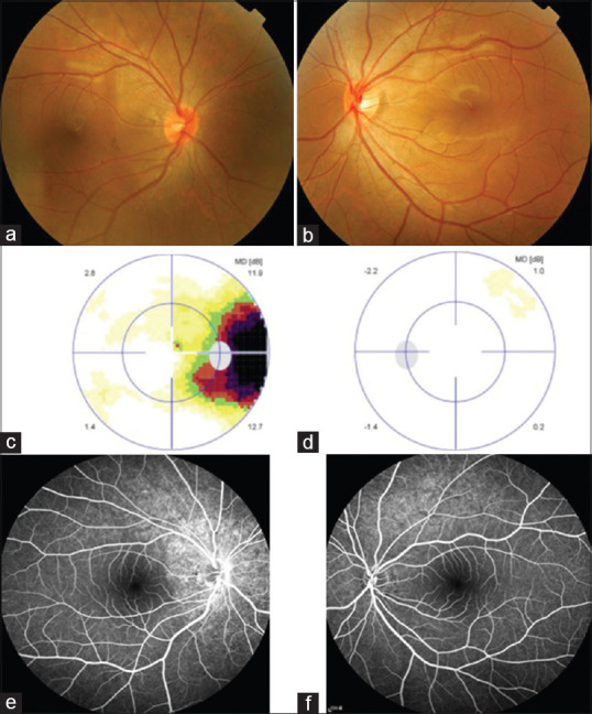
Acute zonal occult outer retinopathy (AZOOR) is an outer retinal disorder characterized by the acute loss of visual functions. Herein, we report a case of AZOOR presenting features mimicking optic neuritis. A 17-year-old healthy male reported fogginess in the right eye for 2 weeks. His best-corrected visual acuity was 20/20 in both eyes. Results of a color vision test and pupillary reaction were unremarkable. Funduscopic examination revealed a subtle hyperemic disc surrounded by hyperpigmentation in the right eye. Visual field examination confirmed an enlarged blind spot in the affected eye. Fundus autofluorescence imaging revealed zonal hyperautofluorescence around the optic disc. Fluorescein angiography showed optic disc staining and a window defect in the retinal pigment epithelium. Optical coherence tomography demonstrated loss of the ellipsoid line at the corresponding hyperautofluorescent region. All these characteristics indicated a diagnosis of AZOOR. However, the prolonged P100 wave observed through visual-evoked potential examination, hyperintensity T2 signal at the retrobulbar optic nerve through magnetic resonance imaging, and mild hyperemic optic disc along with optic disc staining through fluorescein angiography resemble the characteristics of optic neuritis. Because the clinical features of AZOOR are similar to those of optic neuritis, ophthalmologists should be able to differentiate between these two diseases to achieve a timely and correct diagnosis.
急性区域性隐匿性外层视网膜病变(AZOOR)是一种以外层视网膜功能急性丧失为特征的疾病。在此,我们报告一例表现出类似视神经炎特征的AZOOR病例。一名17岁健康男性报告右眼模糊2周。他双眼最佳矫正视力均为20/20。色觉测试和瞳孔反应结果无异常。眼底检查发现右眼视盘轻微充血,周围有色素沉着。视野检查证实患眼盲点扩大。眼底自发荧光成像显示视盘周围有区域性高自发荧光。荧光素血管造影显示视盘染色及视网膜色素上皮窗样缺损。光学相干断层扫描显示相应高自发荧光区域的椭圆体线缺失。所有这些特征均提示AZOOR诊断。然而,视觉诱发电位检查中观察到的P100波延长、磁共振成像显示的球后视神经T2高信号以及荧光素血管造影显示的视盘轻度充血和视盘染色类似于视神经炎的特征。由于AZOOR的临床特征与视神经炎相似,眼科医生应能够区分这两种疾病以便及时、正确诊断。