Duncker Tobias, Lee Winston, Jiang Fan, Ramachandran Rithambara, Hood Donald C, Tsang Stephen H, Sparrow Janet R, Greenstein Vivienne C
Departments of Ophthalmology, and.
Psychology, Columbia University, New York, New York.
Retina. 2018 Jan;38(1):118-127. doi: 10.1097/IAE.0000000000001513.
To assess structure and function across the transition zone (TZ) between relatively healthy and diseased retina in acute zonal occult outer retinopathy.
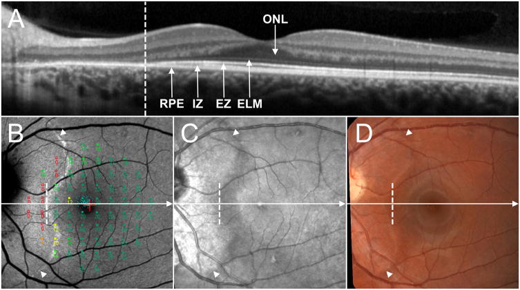
Six patients (6 eyes; age 22-71 years) with acute zonal occult outer retinopathy were studied. Spectral-domain optical coherence tomography, fundus autofluorescence, near-infrared reflectance, color fundus photography, and fundus perimetry were performed and images were registered to each other. The retinal layers of the spectral-domain optical coherence tomography scans were segmented and the thicknesses of two outer retinal layers, that is, the total receptor and outer segment plus layers, and the retinal nerve fiber layer were measured.
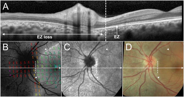
All eyes showed a TZ on multimodal imaging. On spectral-domain optical coherence tomography, the TZ was in the nasal retina at varying distances from the fovea. For all eyes, it was associated with loss of the ellipsoid zone band, significant thinning of the two outer retinal layers, and in three eyes with thickening of the retinal nerve fiber layer. On fundus autofluorescence, all eyes had a clearly demarcated peripapillary area of abnormal fundus autofluorescence delimited by a border of high autofluorescence; the latter was associated with loss of the ellipsoid zone band and with a change from relatively normal to markedly decreased or nonrecordable visual sensitivity on fundus perimetry.
The results of multimodal imaging clarified the TZ in acute zonal occult outer retinopathy. The TZ was outlined by a distinct high autofluorescence border that correlated with loss of the ellipsoid zone band on spectral-domain optical coherence tomography. However, in fundus areas that seemed healthy on fundus autofluorescence, thinning of the outer retinal layers and thickening of the retinal nerve fiber layer were observed near the TZ. The TZ was also characterized by a decrease in visual sensitivity.
评估急性区域性隐匿性外层视网膜病变中相对健康与患病视网膜之间过渡区(TZ)的结构和功能。
对6例(6只眼;年龄22 - 71岁)急性区域性隐匿性外层视网膜病变患者进行研究。进行了光谱域光学相干断层扫描、眼底自发荧光、近红外反射、彩色眼底照相和眼底视野检查,并将图像相互配准。对光谱域光学相干断层扫描图像的视网膜各层进行分割,测量两个外层视网膜层(即总感受器层和外节加层)以及视网膜神经纤维层的厚度。
所有患眼在多模态成像上均显示出一个过渡区。在光谱域光学相干断层扫描上,过渡区位于鼻侧视网膜,距黄斑中心凹距离不等。所有患眼中,过渡区均与椭圆体带缺失、两个外层视网膜层显著变薄相关,3只患眼还与视网膜神经纤维层增厚相关。在眼底自发荧光检查中,所有患眼均有一个以高自发荧光边界界定的清晰划定的视乳头周围异常眼底自发荧光区域;后者与椭圆体带缺失以及眼底视野检查中视觉敏感度从相对正常变为显著降低或无法记录相关。
多模态成像结果明确了急性区域性隐匿性外层视网膜病变中的过渡区。过渡区由一条明显的高自发荧光边界勾勒,该边界与光谱域光学相干断层扫描上椭圆体带缺失相关。然而,在眼底自发荧光看起来正常的区域,在过渡区附近观察到外层视网膜层变薄和视网膜神经纤维层增厚。过渡区还具有视觉敏感度降低的特征。