Shaw Robert C, Tamagnan Gilles D, Tavares Adriana Alexandre S
BHF Centre for Cardiovascular Sciences, University of Edinburgh, Edinburgh, United Kingdom.
Edinburgh Imaging, University of Edinburgh, Edinburgh, United Kingdom.
Front Neurosci. 2020 Oct 1;14:871. doi: 10.3389/fnins.2020.00871. eCollection 2020.
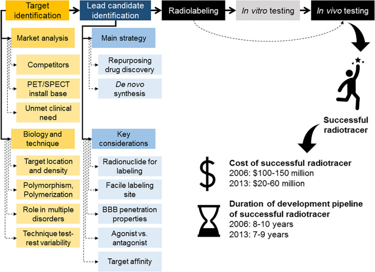
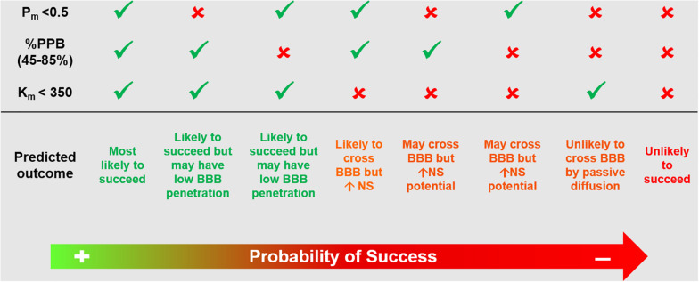
The advent of preclinical research scanners for imaging of small animals has added confidence into the multi-step decision-making process of radiotracer discovery and development. Furthermore, it has expanded the utility of imaging techniques available to dissect clinical questions, fostering a cyclic interaction between the clinical and the preclinical worlds. Significant efforts from medicinal chemistry have also made available several high-affinity and selective compounds amenable for radiolabeling, that target different receptors, transporters and enzymes . This substantially increased the range of applications of molecular imaging using positron emission tomography (PET) or single photon emission computed tomography (SPECT). However, the process of developing novel radiotracers for imaging of the human brain is a multi-step process that has several inherent pitfalls and technical difficulties, which often hampers the successful translation of novel imaging agents from preclinical research into clinical use. In this paper, the process of radiotracer development and its relevance in brain research is discussed; as well as, its pitfalls, technical challenges and future promises. Examples of successful and unsuccessful translation of brain radiotracers will be presented.
用于小动物成像的临床前研究扫描仪的出现,为放射性示踪剂的发现和开发这一多步骤决策过程增添了信心。此外,它还扩展了用于剖析临床问题的成像技术的效用,促进了临床和临床前领域之间的循环互动。药物化学领域的大量努力也提供了几种适用于放射性标记的高亲和力和选择性化合物,这些化合物靶向不同的受体、转运体和酶。这极大地增加了使用正电子发射断层扫描(PET)或单光子发射计算机断层扫描(SPECT)进行分子成像的应用范围。然而,开发用于人脑成像的新型放射性示踪剂的过程是一个多步骤过程,存在一些固有的陷阱和技术难题,这常常阻碍新型成像剂从临床前研究成功转化为临床应用。本文讨论了放射性示踪剂的开发过程及其在脑研究中的相关性;以及其陷阱、技术挑战和未来前景。还将介绍脑放射性示踪剂成功和不成功转化的实例。