Department of Veterinary Microbiology and Pathology, Washington State University, Pullman, WA, 99164, USA.
Division of Infectious Diseases, Department of Medicine, Washington University School of Medicine, St. Louis, MO, 63110, USA.
Int J Parasitol Drugs Drug Resist. 2020 Dec;14:167-182. doi: 10.1016/j.ijpddr.2020.10.007. Epub 2020 Oct 20.
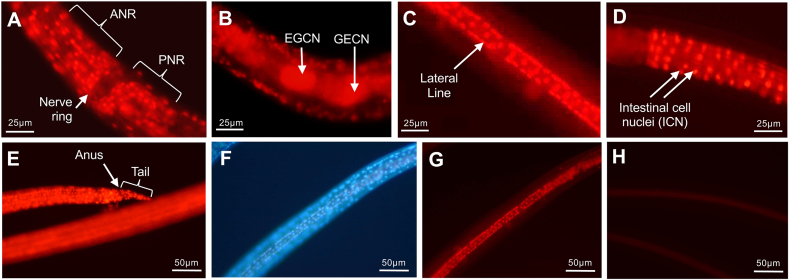
In research focused on the intestine of parasitic nematodes, we recently identified small molecule inhibitors toxic to intestinal cells of larval Ascaris suum (nematode intestinal toxins/toxicants; "NITs"). Some NITs had anthelmintic activity across the phylogenetic diversity of the Nematoda. The whole-worm motility inhibition assay quantified anthelmintic activity, but worm responses to NITs in relation to pathology or affected molecular pathways was not acquired. In this study we extended this research to more comprehensively determine in whole larval A. suum the cells, organ systems, molecular targets, and potential cellular pathways involved in mechanisms of toxicity leading to cell death. The experimental system utilized fluorescent nuclear probes (bisbenzimide, propidium iodide), NITs, an A. suum larval parasite culture system and transcriptional responses (RNA-seq) to NITs. The approach provides for rapid resolution of NIT-induced cell death among organ systems (e.g. intestine, excretory, esophagus, hypodermis and seam cells, and nervous), discriminates among NITs based on cell death profiles, and identifies cells and organ systems with the greatest NIT sensitivity (e.g. intestine and apparent neuronal cells adjacent to the nerve ring). Application was extended to identify cells and organs sensitive to several existing anthelmintics. This approach also resolved intestinal cell death and irreparable damage induced in adult A. suum by two NITs, establishing a new model to elucidate relevant pathologic mechanisms in adult worms. RNA-seq analysis resolved A. suum genes responsive to treatments with three NITs, identifying dihydroorotate dehydrogenase (uridine synthesis) and RAB GTPase(s) (vesicle transport) as potential targets/pathways leading to cell death. A set of genes induced by all three NITs tested suggest common stress or survival responses activated by NITs. Beyond the presented specific lines of research, elements of the overall experimental system presented in this study have broad application toward systematic development of new anthelmintics.
在专注于寄生性线虫肠道的研究中,我们最近鉴定出了一些小分子抑制剂,这些抑制剂对猪蛔虫(Ascaris suum)幼虫的肠道细胞有毒性(线虫肠道毒素/毒物;“NITs”)。一些 NITs 在整个线虫的系统发育多样性中都具有驱虫活性。整体蠕虫运动抑制测定法量化了驱虫活性,但未获得蠕虫对 NITs 的反应与病理学或受影响的分子途径之间的关系。在这项研究中,我们进一步扩展了这项研究,以更全面地确定在整个猪蛔虫幼虫中,参与导致细胞死亡的毒性机制的细胞、器官系统、分子靶标和潜在的细胞途径。该实验系统利用荧光核探针(双苯并咪唑、碘化丙啶)、NITs、猪蛔虫幼虫寄生虫培养系统和转录反应(RNA-seq)对 NITs 的反应。该方法可快速解析 NIT 诱导的器官系统(例如肠道、排泄、食管、皮下组织和 seam 细胞以及神经)之间的细胞死亡,根据细胞死亡谱区分 NITs,并确定对 NIT 最敏感的细胞和器官系统(例如肠道和紧邻神经环的明显神经元细胞)。该方法还扩展到识别对几种现有驱虫药敏感的细胞和器官。该方法还解析了两种 NIT 诱导的成年猪蛔虫肠道细胞死亡和不可修复的损伤,建立了一种新的模型来阐明成年蠕虫中相关的病理机制。RNA-seq 分析解析了对三种 NIT 处理有反应的猪蛔虫基因,确定二氢乳清酸脱氢酶(尿嘧啶合成)和 RAB GTPase(囊泡运输)作为导致细胞死亡的潜在靶标/途径。所有三种 NIT 测试诱导的一组基因表明,NITs 激活了共同的应激或生存反应。除了提出的具体研究方向外,本研究中提出的总体实验系统的各个要素在系统地开发新驱虫药方面具有广泛的应用。