Kulkeaw Kasem, Tubsuwan Alisa, Tongkrajang Nongnat, Whangviboonkij Narisara
Department of Parasitology, Faculty of Medicine Siriraj Hospital, Mahidol University, Bangkok, Thailand.
Stem Cell Research Group, Institute of Molecular Biosciences, Mahidol University, Nakhon Pathom, Thailand.
PeerJ. 2020 Oct 19;8:e9968. doi: 10.7717/peerj.9968. eCollection 2020.
The use of a personalized liver organoid derived from human-induced pluripotent stem cells (HuiPSCs) is advancing the use of in vitro disease models for the design of specific, effective therapies for individuals. Collecting patient peripheral blood cells for HuiPSC generation is preferable because it is less invasive; however, the capability of blood cell-derived HuiPSCs for hepatic differentiation and liver organoid formation remains uncertain. Moreover, the currently available methods for liver organoid formation require a multistep process of cell differentiation or a combination of hepatic endodermal, endothelial and mesenchymal cells, which is a major hurdle for the application of personalized liver organoids in high-throughput testing of drug toxicity and safety. To demonstrate the capability of blood cell-derived HuiPSCs for liver organoid formation without support from endothelial and mesenchymal cells.
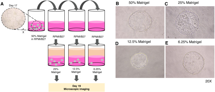
The peripheral blood-derived HuiPSCs first differentiated into hepatic endoderm (HE) in two-dimensional (2D) culture on Matrigel-coated plates under hypoxia for 10 days. The HE was then collected and cultured in 3D culture using 50% Matrigel under ambient oxygen. The maturation of hepatocytes was further induced by adding hepatocyte growth medium containing HGF and oncostatin M on top of the 3D culture and incubating the culture for an additional 12-17 days. The function of the liver organoids was assessed using expression analysis of hepatocyte-specific gene and proteins. Albumin (ALB) synthesis, glycogen and lipid storage, and metabolism of indocyanine were evaluated. The spatial distribution of albumin was examined using immunofluorescence and confocal microscopy.
CD34+ hematopoietic cell-derived HuiPSCs were capable of differentiating into definitive endoderm expressing and , hepatic endoderm expressing , hepatoblasts expressing and hepatocytes expressing . On day 25 of the 2D culture, cells expressed , , and , indicating the presence of cellular heterogeneity. In contrast, the hepatic endoderm spontaneously formed a spherical, hollow structure in a 3D culture of 50% Matrigel, whereas hepatoblasts and hepatocytes could not form. Microscopic observation showed a single layer of polygonal-shaped cells arranged in a 3D structure. The hepatic endoderm-derived organoid synthesis ALB at a higher level than the 2D culture but did not express definitive endoderm-specific , indicating the greater maturity of the hepatocytes in the liver organoids. Confocal microscopic images and quantitative ELISA confirmed albumin synthesis in the cytoplasm of the liver organoid and its secretion. Overall, 3D culture of the hepatic endoderm is a relatively fast, simple, and less laborious way to generate liver organoids from HuiPSCs that is more physiologically relevant than 2D culture.

使用源自人诱导多能干细胞(HuiPSCs)的个性化肝类器官正在推动体外疾病模型的应用,以设计针对个体的特异性、有效疗法。收集患者外周血细胞用于生成HuiPSCs较为可取,因为其侵入性较小;然而,血细胞来源的HuiPSCs向肝分化及形成肝类器官的能力仍不确定。此外,目前可用的肝类器官形成方法需要细胞分化的多步骤过程或肝内胚层、内皮细胞和间充质细胞的组合,这是个性化肝类器官在药物毒性和安全性高通量测试中应用的主要障碍。为了证明血细胞来源的HuiPSCs在无内皮细胞和间充质细胞支持下形成肝类器官的能力。
外周血来源的HuiPSCs首先在涂有基质胶的平板上于缺氧条件下进行二维(2D)培养10天,分化为肝内胚层(HE)。然后收集HE并在含50%基质胶的三维(3D)培养中于环境氧条件下培养。通过在3D培养物顶部添加含肝细胞生长因子(HGF)和制瘤素M的肝细胞生长培养基并再培养12 - 17天,进一步诱导肝细胞成熟。使用肝细胞特异性基因和蛋白质的表达分析评估肝类器官的功能。评估白蛋白(ALB)合成、糖原和脂质储存以及吲哚菁绿代谢。使用免疫荧光和共聚焦显微镜检查白蛋白的空间分布。
CD34 +造血细胞来源的HuiPSCs能够分化为表达 和 的定形内胚层、表达 的肝内胚层、表达 和 的肝母细胞以及表达 的肝细胞。在2D培养的第25天,细胞表达 、 、 和 ,表明存在细胞异质性。相比之下,肝内胚层在含50%基质胶的3D培养中自发形成球形中空结构,而肝母细胞和肝细胞无法形成。显微镜观察显示单层多边形细胞排列成3D结构。肝内胚层来源的类器官合成ALB的水平高于2D培养,但不表达定形内胚层特异性 ,表明肝类器官中肝细胞的成熟度更高。共聚焦显微镜图像和定量酶联免疫吸附测定(ELISA)证实肝类器官细胞质中白蛋白的合成及其分泌。总体而言,肝内胚层的3D培养是一种相对快速、简单且省力的从HuiPSCs生成肝类器官的方法,比2D培养在生理上更相关。