Wang Gongming, Lin Yongyong, Zheng Lie, Liang Yin, Zhao Lei, Wen Yinsheng, Zhang Rusi, Huang Zirui, Yang Longjun, Zhao Dechang, Lachkar Samy, Baste Jean Marc, Shinagawa Naofumi, Ng Calvin S H, Sato Masaaki, Kim Min P, Zhang Lanjun
Department of Thoracic Surgery, Sun Yat-sen University Cancer Center, Guangzhou, China.
Department of Pulmonology, Thoracic Oncology and Respiratory Intensive Care & CIC- CRB 1404, Rouen University Hospital, Rouen, France.
J Thorac Dis. 2020 Sep;12(9):4973-4984. doi: 10.21037/jtd-20-2089.
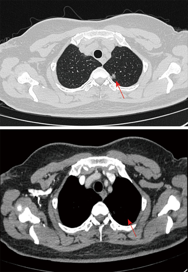
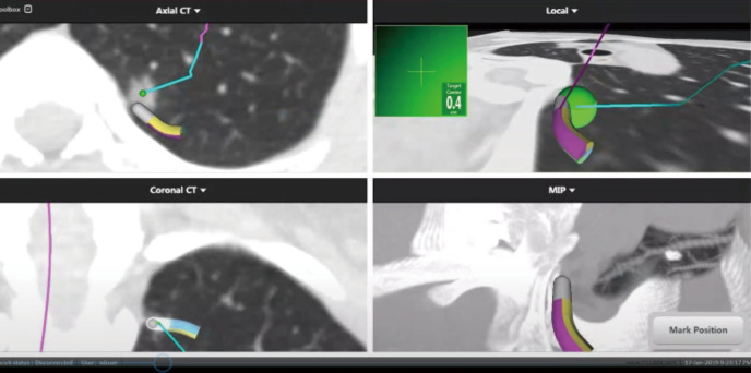
With the use of low-dose CT for early screening of lung cancer, more and more early lung cancers are found. At the same time, patients with small lung nodules have also increased, it is a great challenge for surgeons to resect pulmonary nodules with small volume, deep position and no solid components under video-assisted thoracoscopic surgery. Many studies have reported preoperative and intraoperative methods for localizing lung nodules before minimally invasive resection. Methods for preoperative localization include CT-guided hook-wire positioning, coil positioning, or dye injection and radionuclide location Methods for intraoperative localization include intraoperative ultrasound localization and tactile pressure-sensing localization. After the localization of pulmonary nodules under the guidance of CT patients need to restrict their activities; otherwise, it is easy for the nodules to move, causing the operation to fail, and may also cause complications such as pneumothorax, puncture site pain, and pulmonary parenchymal bleeding. In the past, we injected melamine dye under the guidance of electromagnetic navigation bronchoscope to locate lung nodules. The purpose of this case is introducing a new method for accurately localizing and resecting pulmonary nodules by injecting indocyanine green (ICG) under the guidance of electromagnetic navigation bronchoscope and the resection of small pulmonary nodules under the fluoroscope.
随着低剂量CT用于肺癌早期筛查,越来越多的早期肺癌被发现。与此同时,肺小结节患者也增多了,对于外科医生来说,在电视辅助胸腔镜手术下切除体积小、位置深且无实性成分的肺结节是一项巨大挑战。许多研究报道了微创切除术前和术中定位肺结节的方法。术前定位方法包括CT引导下的钩丝定位、线圈定位、染料注射或放射性核素定位;术中定位方法包括术中超声定位和触觉压力传感定位。在CT引导下对肺结节进行定位后,患者需要限制活动;否则,结节很容易移动,导致手术失败,还可能引起气胸、穿刺部位疼痛和肺实质出血等并发症。过去,我们在电磁导航支气管镜引导下注射三聚氰胺染料来定位肺结节。本病例的目的是介绍一种在电磁导航支气管镜引导下注射吲哚菁绿(ICG)精确地定位和切除肺结节以及在荧光镜下切除小肺结节的新方法。