Qiu Ting, Cui Lei, Xu Jian-Jiang, Hong Jia-Xu, Xiang Jun
Department of Ophthalmology, Shanghai Children's Medical Center, Shanghai Jiao Tong University School of Medicine, Shanghai, China.
National Engineering Research Center for Tissue Engineering, Shanghai, China.
Ann Transl Med. 2020 Sep;8(17):1062. doi: 10.21037/atm-20-4368.
Corneal disease is the second most common cause of blindness in China. Clinically, treatment options for corneal diseases with limbal stem cell deficiency (LSCD) are limited due to a shortage of organ donors and inevitable immune rejection. This study aims to determine the efficacy of reconstructing the ocular surface using autologous cultivated adipose tissue-derived stem cells (ADSCs) and to develop a new clinical treatment for patients with LSCD.
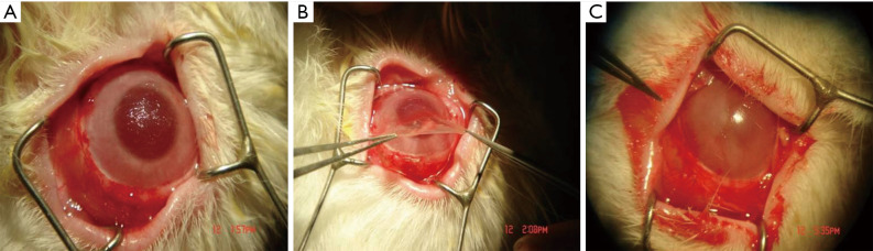
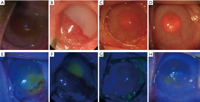
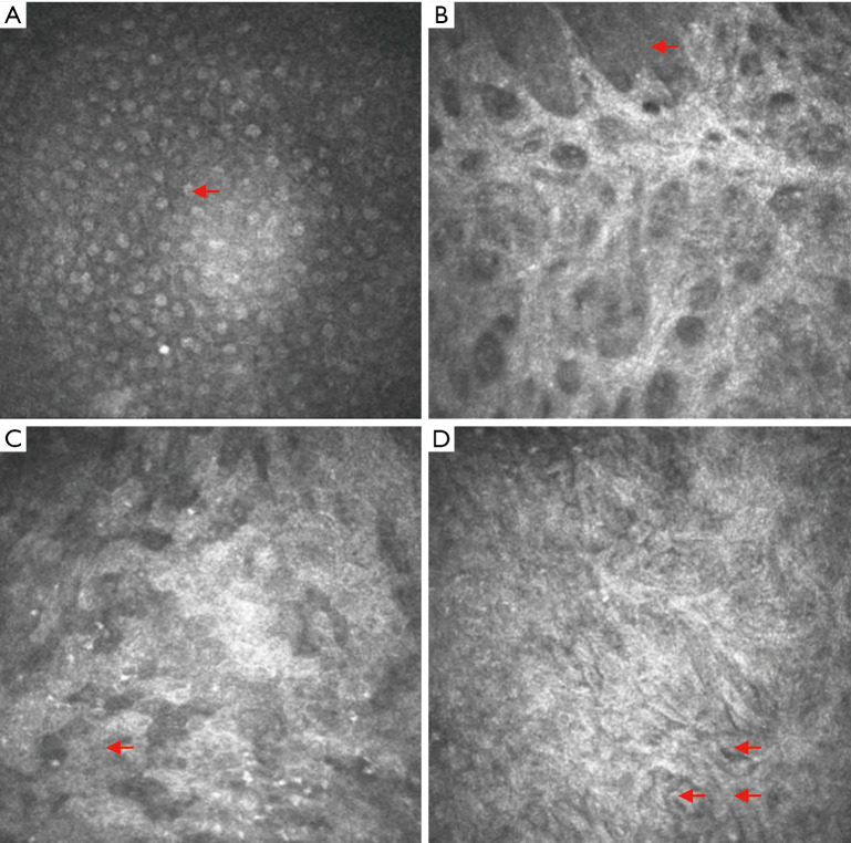
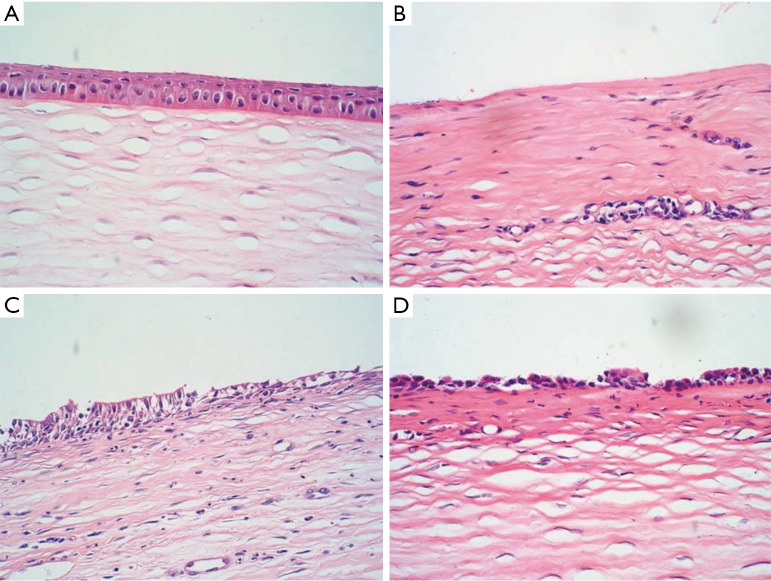
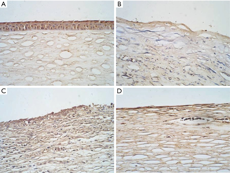
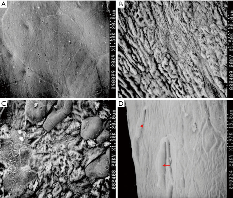
A rabbit LSCD model was first established. Two weeks later, the animals were divided into three groups, including the sham group, the amniotic membrane transplantation group, and the ADSC combined with amniotic membrane transplantation group, and underwent surgery. The efficacy of reconstructing the ocular surface using ADSCs was evaluated using immunofluorescent staining, confocal microscopy (CM) observation, H&E staining, immunohistochemical staining, and scanning transmission electron microscopy observation one, two and four weeks after surgery.
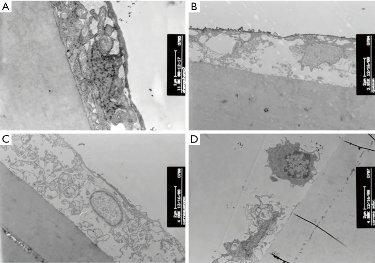
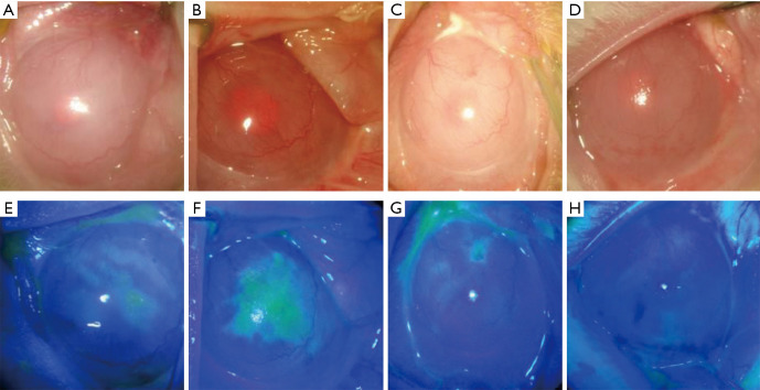
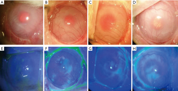
Evaluations of immunofluorescent staining of the cornea pre- and post-surgery yielded significantly lower scores for the corneas in the ADSCs transplantation group than for those in the sham group (F=-7, P=0.002, <0.05) and the amniotic membrane transplantation group (F=-4.67, P=0.018, <0.05) two weeks after surgery. Four weeks after surgery, the corneas of the ADSC combined with amniotic membrane transplantation group were scored significantly lower than those in the sham group (F=-8, P=0.007, <0.05) and the amniotic membrane transplantation group (F=-5.33, P=0.046, <0.05). The data suggest that the use of ADSCs to treat LSCD showed greater efficacy than the other treatment methods. The growth of ADSCs on the corneal surface was examined using confocal and electron microscopes. K3/K12 expression in the corneal epithelium, which was reconstructed by ADSCs, was negative, as shown by immunohistochemical staining.
Ocular surface reconstruction can be improved by using ADSCs as seed cells and the amniotic membrane as a carrier, thus providing a new therapeutic strategy for patients with LSCD.
角膜疾病是中国第二大致盲原因。临床上,由于器官供体短缺和不可避免的免疫排斥反应,针对伴有角膜缘干细胞缺乏(LSCD)的角膜疾病的治疗选择有限。本研究旨在确定使用自体培养的脂肪组织来源干细胞(ADSCs)重建眼表的疗效,并为LSCD患者开发一种新的临床治疗方法。
首先建立兔LSCD模型。两周后,将动物分为三组,包括假手术组、羊膜移植组和ADSC联合羊膜移植组,并进行手术。在术后1周、2周和4周,通过免疫荧光染色、共聚焦显微镜(CM)观察、苏木精-伊红(H&E)染色、免疫组织化学染色和扫描透射电子显微镜观察,评估使用ADSCs重建眼表的疗效。
术后2周,ADSCs移植组角膜免疫荧光染色评分显著低于假手术组(F = -7,P = 0.002,<0.05)和羊膜移植组(F = -4.67,P = 0.018,<0.05)。术后4周,ADSC联合羊膜移植组角膜评分显著低于假手术组(F = -8,P = 0.007,<0.05)和羊膜移植组(F = -5.33,P = 0.046,<0.05)。数据表明,使用ADSCs治疗LSCD比其他治疗方法显示出更高的疗效。使用共聚焦显微镜和电子显微镜检查了ADSCs在角膜表面的生长情况。免疫组织化学染色显示,由ADSCs重建的角膜上皮中K3/K12表达为阴性。
以ADSCs为种子细胞、羊膜为载体可改善眼表重建,从而为LSCD患者提供一种新的治疗策略。