Department of Health Science, Pathology and Anatomy, Hamamatsu University School of Medicine, Hamamatsu, Japan.
PLoS One. 2020 Nov 4;15(11):e0234759. doi: 10.1371/journal.pone.0234759. eCollection 2020.
Aging causes stiffness and decreased function of the renal artery (RA). Histological study with light microscopy can reveal microscopic structural remodeling but no functional changes. The present study aimed to clarify the association between structural and functional aging of the RA through the use of scanning acoustic microscopy.
Formalin-fixed, paraffin-embedded cross-sections of renal arteries from 64 autopsy cases were examined. Speed-of-sound (SOS) values of three layers, which correspond to the stiffness, were compared among different age groups. SOS of the tunica media was examined in terms of blood pressure (BP) and SOS of the ascending aorta. Vulnerability to proteases was assessed by SOS reduction after collagenase treatment.
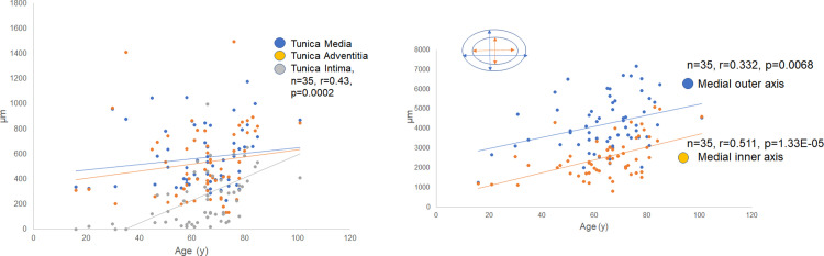
The tunica intima presented inward hypertrophy with luminal narrowing, and the tunica media showed outward hypertrophic remodeling with aging. SOS of the tunica media and internal and external elastic laminae showed a reverse correlation with age. SOS of the tunica media was negatively correlated with BP and strongly associated with that of the aorta. The tunica media of young RAs were more sensitive to collagenase compared with the old ones.
Scanning acoustic microscopy is useful for observing the aging process of the RA. This technique simultaneously shows structural and mechanical information from each portion of the RA. In the process of aging, the RA loses contractile function and elasticity as a result of protease digestion. The tunica media and the internal and external elastic laminae exhibit reduced stiffness, but the tunica intima stiffens with atherosclerosis. As a consequence, the RA's outer shape changes from round to oval with inward and outward hypertrophy. This indicates that the inner resistant intima supports the mechanical weakness of the tunica media to compensate for an increase in BP with aging.
衰老会导致肾动脉(RA)僵硬和功能下降。组织学研究中的光学显微镜检查可以揭示微观结构重塑,但不能显示功能变化。本研究旨在通过使用扫描声学显微镜来阐明 RA 的结构和功能老化之间的关联。
对 64 例尸检肾动脉的福尔马林固定、石蜡包埋的横断面进行检查。比较了不同年龄组三个层(分别对应僵硬程度)的声速(SOS)值。从血压(BP)和升主动脉的 SOS 方面检查中膜的 SOS。通过胶原酶处理后 SOS 降低来评估对蛋白酶的易感性。
内膜呈现向内腔的肥厚性狭窄,中膜呈现向外的肥厚性重塑。中膜的 SOS 与年龄呈反向相关。中膜的 SOS 与 BP 呈负相关,与主动脉的 SOS 密切相关。与老年 RA 相比,年轻 RA 的中膜对胶原酶更敏感。
扫描声学显微镜可用于观察 RA 的老化过程。该技术可同时显示 RA 各部分的结构和机械信息。在老化过程中,RA 由于蛋白酶消化而失去收缩功能和弹性。中膜和内外弹性层的刚性降低,但动脉粥样硬化使内膜变硬。结果,RA 的外形从圆形变为椭圆形,出现向内腔和外腔的肥厚。这表明,具有抗性的内膜支撑着中膜的机械弱点,以补偿随年龄增长而增加的 BP。