Institute of Imaging and Computer Vision, RWTH Aachen University, Aachen, Germany.
Institute of Pathology, RWTH Aachen University Hospital, Aachen, Germany.
J Am Soc Nephrol. 2021 Jan;32(1):52-68. doi: 10.1681/ASN.2020050597. Epub 2020 Nov 5.
Nephropathologic analyses provide important outcomes-related data in experiments with the animal models that are essential for understanding kidney disease pathophysiology. Precision medicine increases the demand for quantitative, unbiased, reproducible, and efficient histopathologic analyses, which will require novel high-throughput tools. A deep learning technique, the convolutional neural network, is increasingly applied in pathology because of its high performance in tasks like histology segmentation.
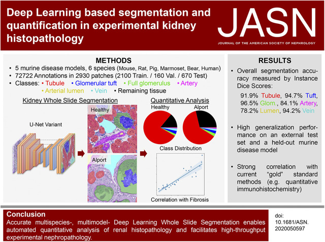
We investigated use of a convolutional neural network architecture for accurate segmentation of periodic acid-Schiff-stained kidney tissue from healthy mice and five murine disease models and from other species used in preclinical research. We trained the convolutional neural network to segment six major renal structures: glomerular tuft, glomerulus including Bowman's capsule, tubules, arteries, arterial lumina, and veins. To achieve high accuracy, we performed a large number of expert-based annotations, 72,722 in total.
Multiclass segmentation performance was very high in all disease models. The convolutional neural network allowed high-throughput and large-scale, quantitative and comparative analyses of various models. In disease models, computational feature extraction revealed interstitial expansion, tubular dilation and atrophy, and glomerular size variability. Validation showed a high correlation of findings with current standard morphometric analysis. The convolutional neural network also showed high performance in other species used in research-including rats, pigs, bears, and marmosets-as well as in humans, providing a translational bridge between preclinical and clinical studies.
We developed a deep learning algorithm for accurate multiclass segmentation of digital whole-slide images of periodic acid-Schiff-stained kidneys from various species and renal disease models. This enables reproducible quantitative histopathologic analyses in preclinical models that also might be applicable to clinical studies.
肾病理分析为理解肾脏疾病病理生理学提供了重要的与结果相关的数据,这些数据对于动物模型的实验至关重要。精准医学增加了对定量、客观、可重复和高效的组织病理学分析的需求,这将需要新的高通量工具。卷积神经网络是一种深度学习技术,由于其在组织学分割等任务中的高性能,在病理学中得到了越来越多的应用。
我们研究了卷积神经网络架构在准确分割来自健康小鼠和五种小鼠疾病模型以及用于临床前研究的其他物种的过碘酸雪夫氏染色肾脏组织中的应用。我们训练卷积神经网络分割六个主要的肾脏结构:肾小球簇、包括鲍曼氏囊的肾小球、肾小管、动脉、动脉管腔和静脉。为了达到高精度,我们进行了大量的基于专家的注释,总共 72722 个。
在所有疾病模型中,多类分割性能都非常高。卷积神经网络允许高通量和大规模、定量和比较分析各种模型。在疾病模型中,计算特征提取揭示了间质扩张、管状扩张和萎缩以及肾小球大小变异性。验证表明,该发现与当前标准形态计量分析具有高度相关性。卷积神经网络在其他用于研究的物种中也表现出了很高的性能,包括大鼠、猪、熊和狨猴,以及人类,为临床前和临床研究之间提供了一个转化桥梁。
我们开发了一种深度学习算法,用于准确分割来自不同物种和肾脏疾病模型的过碘酸雪夫氏染色肾脏数字全幻灯片图像的多类。这使得在临床前模型中进行可重复的定量组织病理学分析成为可能,并且也可能适用于临床研究。