• 文献检索
  • 文档翻译
  • 深度研究
  • 学术资讯
  • Suppr Zotero 插件Zotero 插件
  • 邀请有礼
  • 套餐&价格
  • 历史记录
应用&插件
Suppr Zotero 插件Zotero 插件浏览器插件Mac 客户端Windows 客户端微信小程序
定价
高级版会员购买积分包购买API积分包
服务
文献检索文档翻译深度研究API 文档MCP 服务
关于我们
关于 Suppr公司介绍联系我们用户协议隐私条款
关注我们

Suppr 超能文献

核心技术专利:CN118964589B侵权必究
粤ICP备2023148730 号-1Suppr @ 2026

文献检索

告别复杂PubMed语法,用中文像聊天一样搜索,搜遍4000万医学文献。AI智能推荐,让科研检索更轻松。

立即免费搜索

文件翻译

保留排版,准确专业,支持PDF/Word/PPT等文件格式,支持 12+语言互译。

免费翻译文档

深度研究

AI帮你快速写综述,25分钟生成高质量综述,智能提取关键信息,辅助科研写作。

立即免费体验

肺水肿:影像学表现及疾病机制的当前认识的图文综述

Pulmonary Edema: A Pictorial Review of Imaging Manifestations and Current Understanding of Mechanisms of Disease.

作者信息

Barile Maria

机构信息

Department of Radiology at University of Massachusetts Memorial Medical Center, University of Massachusetts Medical School, Worcester, MA, United States.

出版信息

Eur J Radiol Open. 2020 Oct 30;7:100274. doi: 10.1016/j.ejro.2020.100274. eCollection 2020.

DOI:10.1016/j.ejro.2020.100274
PMID:33163585
原文链接:https://pmc.ncbi.nlm.nih.gov/articles/PMC7607415/
Abstract

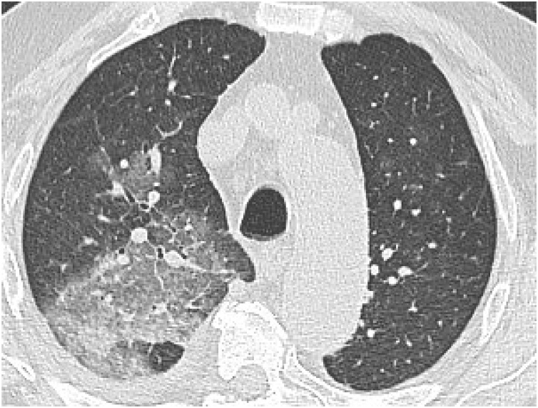
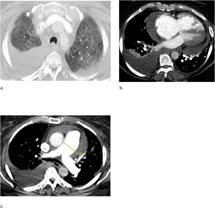
Pulmonary edema is a common clinical entity caused by the extravascular movement of fluid into the pulmonary interstitium and alveoli. The four physiologic categories of edema include hydrostatic pressure edema, permeability edema with and without diffuse alveolar damage (DAD), and mixed edema where there is both an increase in hydrostatic pressure and membrane permeability. As radiographic manifestations and etiologies are varied, an appreciation for both the common and uncommon manifestations and causes of pulmonary edema is essential for accurate diagnosis.

摘要

肺水肿是一种常见的临床病症,由液体向肺间质和肺泡的血管外移动所致。水肿的四种生理类型包括静水压性水肿、伴有或不伴有弥漫性肺泡损伤(DAD)的通透性水肿,以及静水压和膜通透性均增加的混合性水肿。由于影像学表现和病因各不相同,了解肺水肿的常见和不常见表现及病因对于准确诊断至关重要。

https://cdn.ncbi.nlm.nih.gov/pmc/blobs/499b/7607415/5010f8f5d87e/gr20.jpg
https://cdn.ncbi.nlm.nih.gov/pmc/blobs/499b/7607415/857349b90d02/gr1.jpg
https://cdn.ncbi.nlm.nih.gov/pmc/blobs/499b/7607415/5bdba35880f7/gr2.jpg
https://cdn.ncbi.nlm.nih.gov/pmc/blobs/499b/7607415/276fb1c850f9/gr3.jpg
https://cdn.ncbi.nlm.nih.gov/pmc/blobs/499b/7607415/9639af6d4b24/gr4.jpg
https://cdn.ncbi.nlm.nih.gov/pmc/blobs/499b/7607415/3cca3a0bc2c0/gr5.jpg
https://cdn.ncbi.nlm.nih.gov/pmc/blobs/499b/7607415/692415dc8e1b/gr6.jpg
https://cdn.ncbi.nlm.nih.gov/pmc/blobs/499b/7607415/7921e5e637e0/gr7.jpg
https://cdn.ncbi.nlm.nih.gov/pmc/blobs/499b/7607415/98014d35628d/gr8.jpg
https://cdn.ncbi.nlm.nih.gov/pmc/blobs/499b/7607415/e2182ed32e92/gr9.jpg
https://cdn.ncbi.nlm.nih.gov/pmc/blobs/499b/7607415/22a692c240c3/gr10.jpg
https://cdn.ncbi.nlm.nih.gov/pmc/blobs/499b/7607415/32a6f25c8fcc/gr11.jpg
https://cdn.ncbi.nlm.nih.gov/pmc/blobs/499b/7607415/e7f7b00a35f5/gr12.jpg
https://cdn.ncbi.nlm.nih.gov/pmc/blobs/499b/7607415/741cda105003/gr13.jpg
https://cdn.ncbi.nlm.nih.gov/pmc/blobs/499b/7607415/34d34b4152dd/gr14.jpg
https://cdn.ncbi.nlm.nih.gov/pmc/blobs/499b/7607415/2de272106208/gr15.jpg
https://cdn.ncbi.nlm.nih.gov/pmc/blobs/499b/7607415/454f6b4ff1dd/gr16.jpg
https://cdn.ncbi.nlm.nih.gov/pmc/blobs/499b/7607415/94b38baab0a5/gr17.jpg
https://cdn.ncbi.nlm.nih.gov/pmc/blobs/499b/7607415/73d80946adbd/gr18.jpg
https://cdn.ncbi.nlm.nih.gov/pmc/blobs/499b/7607415/50fe49297f44/gr19.jpg
https://cdn.ncbi.nlm.nih.gov/pmc/blobs/499b/7607415/5010f8f5d87e/gr20.jpg

相似文献

1
Pulmonary Edema: A Pictorial Review of Imaging Manifestations and Current Understanding of Mechanisms of Disease.肺水肿:影像学表现及疾病机制的当前认识的图文综述
Eur J Radiol Open. 2020 Oct 30;7:100274. doi: 10.1016/j.ejro.2020.100274. eCollection 2020.
2
A new view of pulmonary edema and acute respiratory distress syndrome.肺水肿与急性呼吸窘迫综合征的新观点
J Thorac Imaging. 1998 Jul;13(3):147-71.
3
Clinical and radiologic features of pulmonary edema.肺水肿的临床和放射学特征。
Radiographics. 1999 Nov-Dec;19(6):1507-31; discussion 1532-3. doi: 10.1148/radiographics.19.6.g99no211507.
4
Assessing pulmonary permeability by transpulmonary thermodilution allows differentiation of hydrostatic pulmonary edema from ALI/ARDS.通过经肺热稀释法评估肺通透性可区分静水压性肺水肿与急性肺损伤/急性呼吸窘迫综合征。
Intensive Care Med. 2007 Mar;33(3):448-53. doi: 10.1007/s00134-006-0498-6. Epub 2007 Jan 13.
5
Noncardiogenic pulmonary edema in small animals.小动物非心源性肺水肿
J Vet Emerg Crit Care (San Antonio). 2023 Mar;33(2):156-172. doi: 10.1111/vec.13278. Epub 2023 Feb 23.
6
The clinical usefulness of extravascular lung water and pulmonary vascular permeability index to diagnose and characterize pulmonary edema: a prospective multicenter study on the quantitative differential diagnostic definition for acute lung injury/acute respiratory distress syndrome.血管外肺水和肺血管通透性指数在诊断和鉴别肺水肿中的临床应用:一项关于急性肺损伤/急性呼吸窘迫综合征定量鉴别诊断定义的前瞻性多中心研究。
Crit Care. 2012 Dec 11;16(6):R232. doi: 10.1186/cc11898.
7
The alveolar space is the site of intense inflammatory and profibrotic reactions in the early phase of acute respiratory distress syndrome.在急性呼吸窘迫综合征的早期阶段,肺泡腔是强烈炎症反应和促纤维化反应的发生部位。
Crit Care Med. 1999 Feb;27(2):304-12. doi: 10.1097/00003246-199902000-00036.
8
Diagnostic value of transpulmonary thermodilution measurements for acute respiratory distress syndrome in a pig model of septic shock.经肺热稀释测量对感染性休克猪模型急性呼吸窘迫综合征的诊断价值。
J Transl Med. 2022 Dec 23;20(1):617. doi: 10.1186/s12967-022-03793-x.
9
Pulmonary veno-occlusive disease: clinical, functional, radiologic, and hemodynamic characteristics and outcome of 24 cases confirmed by histology.肺静脉闭塞病:24例经组织学确诊病例的临床、功能、放射学及血流动力学特征与转归
Medicine (Baltimore). 2008 Jul;87(4):220-233. doi: 10.1097/MD.0b013e31818193bb.
10
Distinguishing hantavirus pulmonary syndrome from acute respiratory distress syndrome by chest radiography: are there different radiographic manifestations of increased alveolar permeability?通过胸部X线摄影鉴别汉坦病毒肺综合征与急性呼吸窘迫综合征:肺泡通透性增加是否存在不同的影像学表现?
J Thorac Imaging. 1998 Jul;13(3):172-7. doi: 10.1097/00005382-199807000-00002.

引用本文的文献

1
Quantification of residual pulmonary congestion defined by B-line findings on lung ultrasound to predict cardiovascular events in acute heart failure: A systematic review and meta analysis.通过肺部超声B线表现对残余肺淤血进行量化以预测急性心力衰竭心血管事件:一项系统评价和荟萃分析
Indian Heart J. 2025 Jul-Aug;77(4):258-266. doi: 10.1016/j.ihj.2025.05.003. Epub 2025 May 5.
2
Postextubation negative-pressure pulmonary edema after an appendectomy.阑尾切除术后拔管后负压性肺水肿
Radiol Case Rep. 2025 Mar 31;20(6):3063-3066. doi: 10.1016/j.radcr.2025.03.053. eCollection 2025 Jun.
3
Automated volumetry of core and peel intrapulmonary vasculature on computed tomography angiography for non-invasive estimation of hemodynamics in patients with pulmonary hypertension (2022 updated hemodynamic definition).

本文引用的文献

1
Primary Graft Dysfunction After Lung Transplantation.肺移植后原发性移植物功能障碍。
Clin Chest Med. 2017 Dec;38(4):641-654. doi: 10.1016/j.ccm.2017.07.005. Epub 2017 Sep 20.
2
Report of the ISHLT Working Group on Primary Lung Graft Dysfunction Part III: Mechanisms: A 2016 Consensus Group Statement of the International Society for Heart and Lung Transplantation.国际心肺移植学会原发性肺移植功能障碍工作组报告第三部分:机制——2016年国际心肺移植学会共识组声明
J Heart Lung Transplant. 2017 Oct;36(10):1114-1120. doi: 10.1016/j.healun.2017.07.014. Epub 2017 Jul 24.
3
Orexin activation counteracts decreases in nonexercise activity thermogenesis (NEAT) caused by high-fat diet.
计算机断层血管造影术对肺动脉高压患者肺内核心和外周血管系统进行自动容积测量以无创评估血流动力学(2022年更新的血流动力学定义)
Cardiovasc Diagn Ther. 2024 Dec 31;14(6):1083-1095. doi: 10.21037/cdt-24-293. Epub 2024 Dec 19.
4
Breath-by-breath assessment of acute pulmonary edema using electrical impedance tomography, spirometry and volumetric capnography in a sheep () model.在绵羊模型中使用电阻抗断层成像、肺活量测定法和容积式二氧化碳描记法对急性肺水肿进行逐次呼吸评估。
Front Vet Sci. 2024 Jul 10;11:1402748. doi: 10.3389/fvets.2024.1402748. eCollection 2024.
5
An Assessment of the Prevalence and Risk Factors of Hypertensive Crisis in Patients Who Visited the Emergency Outpatient Department (EOPD) at Adama Hospital Medical College, Adama, Oromia, Ethiopia: A 6-Month Prospective Study.埃塞俄比亚奥罗米亚州阿达马阿达马医院医学院急诊科就诊患者高血压危象患病率及危险因素评估:一项为期6个月的前瞻性研究
Int J Hypertens. 2024 Apr 29;2024:6893267. doi: 10.1155/2024/6893267. eCollection 2024.
6
Risk factors for re-expansion pulmonary edema following chest tube drainage in patients with spontaneous pneumothorax: A systematic review and meta-analysis.自发性气胸患者胸腔闭式引流后复张性肺水肿的危险因素:一项系统评价和荟萃分析
J Cardiovasc Thorac Res. 2024;16(1):1-7. doi: 10.34172/jcvtr.32871. Epub 2024 Mar 13.
7
A deep learning model enables accurate prediction and quantification of pulmonary edema from chest X-rays.深度学习模型可准确预测和量化胸片中的肺水肿。
Crit Care. 2023 May 26;27(1):201. doi: 10.1186/s13054-023-04426-5.
8
Diagnosing Lung Abnormalities Related to Heart Failure in Chest Radiogram, Lung Ultrasound and Thoracic Computed Tomography.诊断与心力衰竭相关的胸部放射影像、肺部超声和胸部计算机断层扫描中的肺部异常。
Adv Respir Med. 2023 Feb 23;91(2):103-122. doi: 10.3390/arm91020010.
9
Lung Dysfunction and Chronic Kidney Disease: A Complex Network of Multiple Interactions.肺功能障碍与慢性肾脏病:多重相互作用的复杂网络
J Pers Med. 2023 Feb 3;13(2):286. doi: 10.3390/jpm13020286.
10
siRNA-Mediated Silencing Inhibited the Inflammatory Phenotype during Acute Lung Injury.siRNA 介导的沉默抑制急性肺损伤中的炎症表型。
Int J Mol Sci. 2023 Jan 13;24(2):1641. doi: 10.3390/ijms24021641.
食欲素激活可抵消高脂饮食引起的非运动活动产热(NEAT)减少。
Physiol Behav. 2017 Jul 1;176:139-148. doi: 10.1016/j.physbeh.2017.03.040. Epub 2017 Mar 28.
4
Pulmonary veno-occlusive disease.肺静脉闭塞病。
Eur Respir J. 2016 May;47(5):1518-34. doi: 10.1183/13993003.00026-2016. Epub 2016 Mar 23.
5
The large spectrum of pulmonary complications following illicit drug use: features and mechanisms.非法药物使用后肺部并发症的广泛谱:特征和机制。
Chem Biol Interact. 2013 Dec 5;206(3):444-51. doi: 10.1016/j.cbi.2013.10.011. Epub 2013 Oct 18.
6
Pulmonary edema predictive scoring index (PEPSI), a new index to predict risk of reperfusion pulmonary edema and improvement of hemodynamics in percutaneous transluminal pulmonary angioplasty.肺充血预测评分指数(PEPSI),一种新的预测经皮腔内肺血管成形术再灌注肺充血风险和改善血液动力学的指标。
JACC Cardiovasc Interv. 2013 Jul;6(7):725-36. doi: 10.1016/j.jcin.2013.03.009. Epub 2013 Jun 14.
7
Neurogenic pulmonary edema.神经源性肺水肿
Crit Care. 2012 Dec 12;16(2):212. doi: 10.1186/cc11226.
8
Post-ARDS pulmonary fibrosis in patients with H1N1 pneumonia: role of follow-up CT.H1N1 肺炎患者并发 ARDS 后肺纤维化:随访 CT 的作用。
Radiol Med. 2012 Mar;117(2):185-200. doi: 10.1007/s11547-011-0740-3. Epub 2011 Oct 21.
9
Reexpansion pulmonary edema after therapeutic thoracentesis.治疗性胸腔穿刺术后复张性肺水肿
Clinics (Sao Paulo). 2010;65(12):1387-9. doi: 10.1590/s1807-59322010001200026.
10
Postoperative complications of lung transplantation: radiologic findings along a time continuum.肺移植术后并发症:随时间推移的影像学表现
Radiographics. 2007 Jul-Aug;27(4):957-74. doi: 10.1148/rg.274065141.