• 文献检索
  • 文档翻译
  • 深度研究
  • 学术资讯
  • Suppr Zotero 插件Zotero 插件
  • 邀请有礼
  • 套餐&价格
  • 历史记录
应用&插件
Suppr Zotero 插件Zotero 插件浏览器插件Mac 客户端Windows 客户端微信小程序
定价
高级版会员购买积分包购买API积分包
服务
文献检索文档翻译深度研究API 文档MCP 服务
关于我们
关于 Suppr公司介绍联系我们用户协议隐私条款
关注我们

Suppr 超能文献

核心技术专利:CN118964589B侵权必究
粤ICP备2023148730 号-1Suppr @ 2026

文献检索

告别复杂PubMed语法,用中文像聊天一样搜索,搜遍4000万医学文献。AI智能推荐,让科研检索更轻松。

立即免费搜索

文件翻译

保留排版,准确专业,支持PDF/Word/PPT等文件格式,支持 12+语言互译。

免费翻译文档

深度研究

AI帮你快速写综述,25分钟生成高质量综述,智能提取关键信息,辅助科研写作。

立即免费体验

用于乳腺组织病理学中可变大小感兴趣区域的深度特征表示。

Deep Feature Representations for Variable-Sized Regions of Interest in Breast Histopathology.

出版信息

IEEE J Biomed Health Inform. 2021 Jun;25(6):2041-2049. doi: 10.1109/JBHI.2020.3036734. Epub 2021 Jun 3.

DOI:10.1109/JBHI.2020.3036734
PMID:33166257
原文链接:https://pmc.ncbi.nlm.nih.gov/articles/PMC8274968/
Abstract

OBJECTIVE

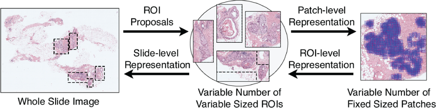
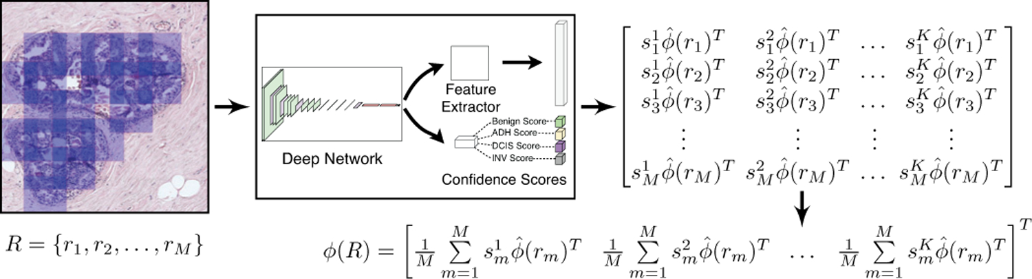
Modeling variable-sized regions of interest (ROIs) in whole slide images using deep convolutional networks is a challenging task, as these networks typically require fixed-sized inputs that should contain sufficient structural and contextual information for classification. We propose a deep feature extraction framework that builds an ROI-level feature representation via weighted aggregation of the representations of variable numbers of fixed-sized patches sampled from nuclei-dense regions in breast histopathology images.

METHODS

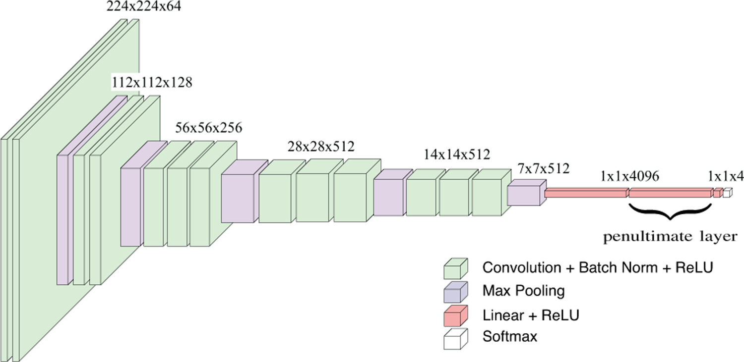
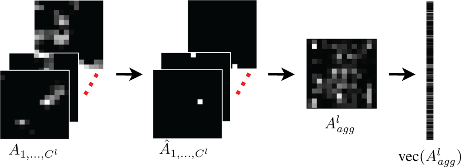
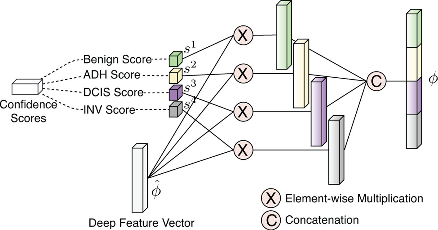
First, the initial patch-level feature representations are extracted from both fully-connected layer activations and pixel-level convolutional layer activations of a deep network, and the weights are obtained from the class predictions of the same network trained on patch samples. Then, the final patch-level feature representations are computed by concatenation of weighted instances of the extracted feature activations. Finally, the ROI-level representation is obtained by fusion of the patch-level representations by average pooling.

RESULTS

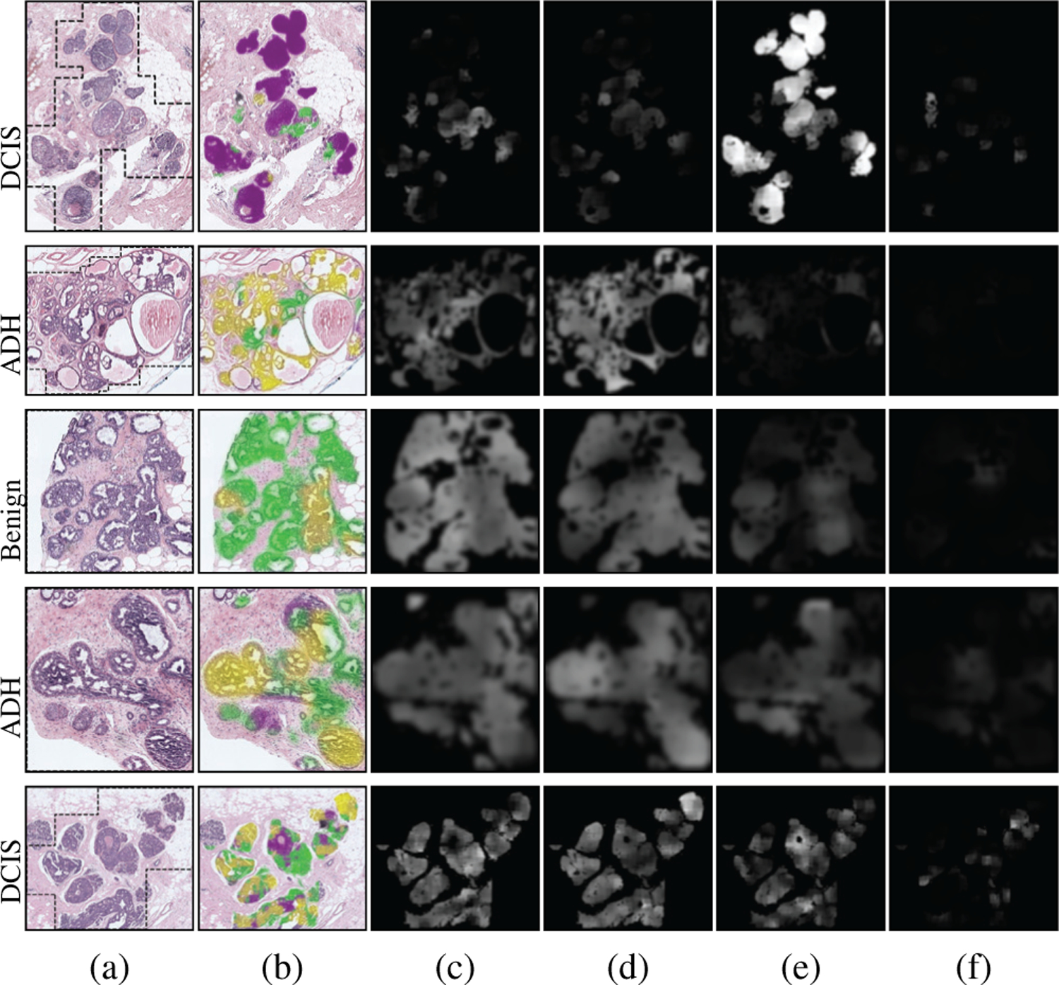
Experiments using a well-characterized data set of 240 slides containing 437 ROIs marked by experienced pathologists with variable sizes and shapes result in an accuracy score of 72.65% in classifying ROIs into four diagnostic categories that cover the whole histologic spectrum.

CONCLUSION

The results show that the proposed feature representations are superior to existing approaches and provide accuracies that are higher than the average accuracy of another set of pathologists.

SIGNIFICANCE

The proposed generic representation that can be extracted from any type of deep convolutional architecture combines the patch appearance information captured by the network activations and the diagnostic relevance predicted by the class-specific scoring of patches for effective modeling of variable-sized ROIs.

摘要

目的

使用深度卷积网络对全切片图像中的可变大小感兴趣区域(ROI)进行建模是一项具有挑战性的任务,因为这些网络通常需要固定大小的输入,这些输入应该包含足够的结构和上下文信息以进行分类。我们提出了一种深度特征提取框架,通过对从乳腺组织病理学图像中核密集区域采样的可变数量的固定大小的斑块的表示进行加权聚合,构建 ROI 级别的特征表示。

方法

首先,从深度网络的全连接层激活和像素级卷积层激活中提取初始斑块级特征表示,并从在斑块样本上训练的相同网络的类别预测中获得权重。然后,通过对提取的特征激活的加权实例进行串联计算最终的斑块级特征表示。最后,通过平均池化融合斑块级表示来获得 ROI 级表示。

结果

使用由经验丰富的病理学家标记的具有可变大小和形状的 240 张幻灯片组成的特征良好的数据集进行的实验,将 ROI 分为四个诊断类别,涵盖整个组织学谱,分类准确率为 72.65%。

结论

结果表明,所提出的特征表示优于现有方法,并提供了高于另一组病理学家平均准确率的准确率。

意义

可以从任何类型的深度卷积架构中提取的这种通用表示形式结合了网络激活捕获的斑块外观信息和斑块特定评分预测的诊断相关性,可有效建模可变大小的 ROI。

https://cdn.ncbi.nlm.nih.gov/pmc/blobs/1fe5/8274968/b2c5908980d7/nihms-1711339-f0008.jpg
https://cdn.ncbi.nlm.nih.gov/pmc/blobs/1fe5/8274968/29aae944e1c9/nihms-1711339-f0001.jpg
https://cdn.ncbi.nlm.nih.gov/pmc/blobs/1fe5/8274968/011f2e839187/nihms-1711339-f0002.jpg
https://cdn.ncbi.nlm.nih.gov/pmc/blobs/1fe5/8274968/2cadbb4a6021/nihms-1711339-f0003.jpg
https://cdn.ncbi.nlm.nih.gov/pmc/blobs/1fe5/8274968/ca4508a0d2c8/nihms-1711339-f0004.jpg
https://cdn.ncbi.nlm.nih.gov/pmc/blobs/1fe5/8274968/c00322b0bc14/nihms-1711339-f0005.jpg
https://cdn.ncbi.nlm.nih.gov/pmc/blobs/1fe5/8274968/52ed4b25c720/nihms-1711339-f0006.jpg
https://cdn.ncbi.nlm.nih.gov/pmc/blobs/1fe5/8274968/74841229ad4b/nihms-1711339-f0007.jpg
https://cdn.ncbi.nlm.nih.gov/pmc/blobs/1fe5/8274968/b2c5908980d7/nihms-1711339-f0008.jpg

相似文献

1
Deep Feature Representations for Variable-Sized Regions of Interest in Breast Histopathology.用于乳腺组织病理学中可变大小感兴趣区域的深度特征表示。
IEEE J Biomed Health Inform. 2021 Jun;25(6):2041-2049. doi: 10.1109/JBHI.2020.3036734. Epub 2021 Jun 3.
2
Computer assisted recognition of breast cancer in biopsy images via fusion of nucleus-guided deep convolutional features.通过细胞核引导的深度卷积特征融合实现活检图像中乳腺癌的计算机辅助识别。
Comput Methods Programs Biomed. 2020 Oct;194:105531. doi: 10.1016/j.cmpb.2020.105531. Epub 2020 May 11.
3
Detection and classification of cancer in whole slide breast histopathology images using deep convolutional networks.使用深度卷积网络对全切片乳腺组织病理学图像中的癌症进行检测和分类。
Pattern Recognit. 2018 Dec;84:345-356. doi: 10.1016/j.patcog.2018.07.022. Epub 2018 Jul 20.
4
Multi-Instance Multi-Label Learning for Multi-Class Classification of Whole Slide Breast Histopathology Images.多实例多标签学习在全切片乳腺组织病理学图像多类分类中的应用。
IEEE Trans Med Imaging. 2018 Jan;37(1):316-325. doi: 10.1109/TMI.2017.2758580. Epub 2017 Oct 2.
5
Mass detection in digital breast tomosynthesis: Deep convolutional neural network with transfer learning from mammography.数字乳腺断层合成中的肿块检测:基于乳腺X线摄影迁移学习的深度卷积神经网络
Med Phys. 2016 Dec;43(12):6654. doi: 10.1118/1.4967345.
6
Cross-Convolutional-Layer Pooling for Image Recognition.跨卷积层池化的图像识别。
IEEE Trans Pattern Anal Mach Intell. 2017 Nov;39(11):2305-2313. doi: 10.1109/TPAMI.2016.2637921. Epub 2016 Dec 9.
7
Breast ultrasound image segmentation: A coarse-to-fine fusion convolutional neural network.乳腺超声图像分割:一种粗到细融合的卷积神经网络。
Med Phys. 2021 Aug;48(8):4262-4278. doi: 10.1002/mp.15006. Epub 2021 Jul 29.
8
Breast cancer detection from biopsy images using nucleus guided transfer learning and belief based fusion.基于核引导迁移学习和置信度融合的活检图像乳腺癌检测
Comput Biol Med. 2020 Sep;124:103954. doi: 10.1016/j.compbiomed.2020.103954. Epub 2020 Aug 4.
9
DV-DCNN: Dual-view deep convolutional neural network for matching detected masses in mammograms.DV-DCNN:用于匹配乳腺X线摄影中检测到的肿块的双视图深度卷积神经网络。
Comput Methods Programs Biomed. 2021 Aug;207:106152. doi: 10.1016/j.cmpb.2021.106152. Epub 2021 May 11.
10
Saliency map-guided hierarchical dense feature aggregation framework for breast lesion classification using ultrasound image.基于显著图引导的层次密集特征聚合框架的超声图像乳腺病变分类方法
Comput Methods Programs Biomed. 2022 Mar;215:106612. doi: 10.1016/j.cmpb.2021.106612. Epub 2021 Dec 31.

引用本文的文献

1
Application of Deep Learning in Histopathology Images of Breast Cancer: A Review.深度学习在乳腺癌组织病理学图像中的应用:综述
Micromachines (Basel). 2022 Dec 11;13(12):2197. doi: 10.3390/mi13122197.
2
Improving the Diagnosis of Skin Biopsies Using Tissue Segmentation.利用组织分割技术改善皮肤活检诊断
Diagnostics (Basel). 2022 Jul 14;12(7):1713. doi: 10.3390/diagnostics12071713.
3
Scale-Aware Transformers for Diagnosing Melanocytic Lesions.用于诊断黑素细胞性病变的尺度感知变压器

本文引用的文献

1
Assessment of Machine Learning of Breast Pathology Structures for Automated Differentiation of Breast Cancer and High-Risk Proliferative Lesions.基于机器学习的乳腺病理学结构评估在乳腺癌和高危增殖性病变自动鉴别中的应用。
JAMA Netw Open. 2019 Aug 2;2(8):e198777. doi: 10.1001/jamanetworkopen.2019.8777.
2
BACH: Grand challenge on breast cancer histology images.BACH:乳腺癌组织学图像的重大挑战。
Med Image Anal. 2019 Aug;56:122-139. doi: 10.1016/j.media.2019.05.010. Epub 2019 May 31.
3
Detection and classification of cancer in whole slide breast histopathology images using deep convolutional networks.
IEEE Access. 2021;9:163526-163541. doi: 10.1109/ACCESS.2021.3132958. Epub 2021 Dec 6.
使用深度卷积网络对全切片乳腺组织病理学图像中的癌症进行检测和分类。
Pattern Recognit. 2018 Dec;84:345-356. doi: 10.1016/j.patcog.2018.07.022. Epub 2018 Jul 20.
4
Patch-based system for Classification of Breast Histology images using deep learning.基于补丁的深度学习系统用于乳腺组织学图像分类。
Comput Med Imaging Graph. 2019 Jan;71:90-103. doi: 10.1016/j.compmedimag.2018.11.003. Epub 2018 Dec 1.
5
Deep Manifold Preserving Autoencoder for Classifying Breast Cancer Histopathological Images.深度流形保持自动编码器在乳腺癌病理图像分类中的应用。
IEEE/ACM Trans Comput Biol Bioinform. 2020 Jan-Feb;17(1):91-101. doi: 10.1109/TCBB.2018.2858763. Epub 2018 Jul 23.
6
Multi-Instance Multi-Label Learning for Multi-Class Classification of Whole Slide Breast Histopathology Images.多实例多标签学习在全切片乳腺组织病理学图像多类分类中的应用。
IEEE Trans Med Imaging. 2018 Jan;37(1):316-325. doi: 10.1109/TMI.2017.2758580. Epub 2017 Oct 2.
7
A survey on deep learning in medical image analysis.深度学习在医学图像分析中的应用研究综述。
Med Image Anal. 2017 Dec;42:60-88. doi: 10.1016/j.media.2017.07.005. Epub 2017 Jul 26.
8
Classification of breast cancer histology images using Convolutional Neural Networks.使用卷积神经网络对乳腺癌组织学图像进行分类
PLoS One. 2017 Jun 1;12(6):e0177544. doi: 10.1371/journal.pone.0177544. eCollection 2017.
9
Accurate and reproducible invasive breast cancer detection in whole-slide images: A Deep Learning approach for quantifying tumor extent.全切片图像中精确且可重复的浸润性乳腺癌检测:一种用于量化肿瘤范围的深度学习方法。
Sci Rep. 2017 Apr 18;7:46450. doi: 10.1038/srep46450.
10
A Randomized Study Comparing Digital Imaging to Traditional Glass Slide Microscopy for Breast Biopsy and Cancer Diagnosis.一项比较数字成像与传统玻璃切片显微镜用于乳腺活检和癌症诊断的随机研究。
J Pathol Inform. 2017 Mar 10;8:12. doi: 10.4103/2153-3539.201920. eCollection 2017.