• 文献检索
  • 文档翻译
  • 深度研究
  • 学术资讯
  • Suppr Zotero 插件Zotero 插件
  • 邀请有礼
  • 套餐&价格
  • 历史记录
应用&插件
Suppr Zotero 插件Zotero 插件浏览器插件Mac 客户端Windows 客户端微信小程序
定价
高级版会员购买积分包购买API积分包
服务
文献检索文档翻译深度研究API 文档MCP 服务
关于我们
关于 Suppr公司介绍联系我们用户协议隐私条款
关注我们

Suppr 超能文献

核心技术专利:CN118964589B侵权必究
粤ICP备2023148730 号-1Suppr @ 2026

文献检索

告别复杂PubMed语法,用中文像聊天一样搜索,搜遍4000万医学文献。AI智能推荐,让科研检索更轻松。

立即免费搜索

文件翻译

保留排版,准确专业,支持PDF/Word/PPT等文件格式,支持 12+语言互译。

免费翻译文档

深度研究

AI帮你快速写综述,25分钟生成高质量综述,智能提取关键信息,辅助科研写作。

立即免费体验

解析人肾纤维化中肌成纤维细胞的起源。

Decoding myofibroblast origins in human kidney fibrosis.

机构信息

Division of Nephrology and Clinical Immunology, RWTH Aachen University, Aachen, Germany.

Institute of Experimental Medicine and Systems Biology, RWTH Aachen University, Aachen, Germany.

出版信息

Nature. 2021 Jan;589(7841):281-286. doi: 10.1038/s41586-020-2941-1. Epub 2020 Nov 11.

DOI:10.1038/s41586-020-2941-1
PMID:33176333
原文链接:https://pmc.ncbi.nlm.nih.gov/articles/PMC7611626/
Abstract

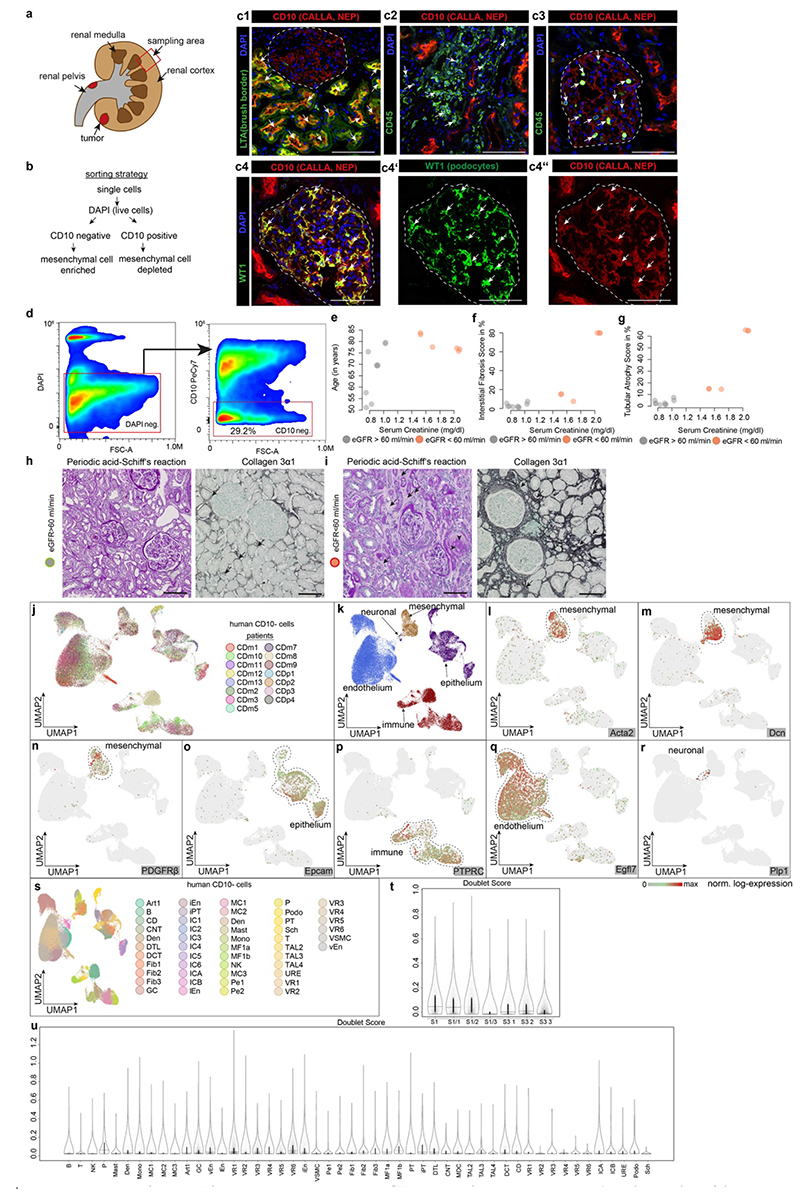
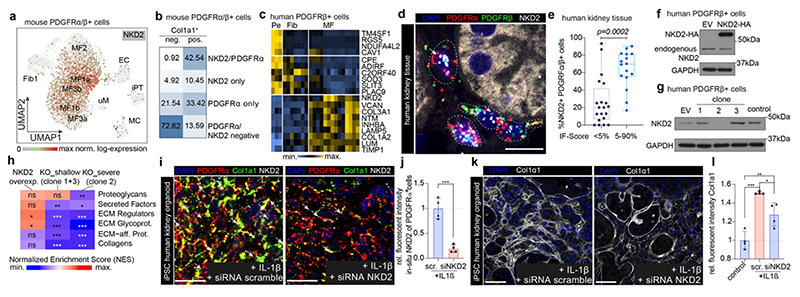
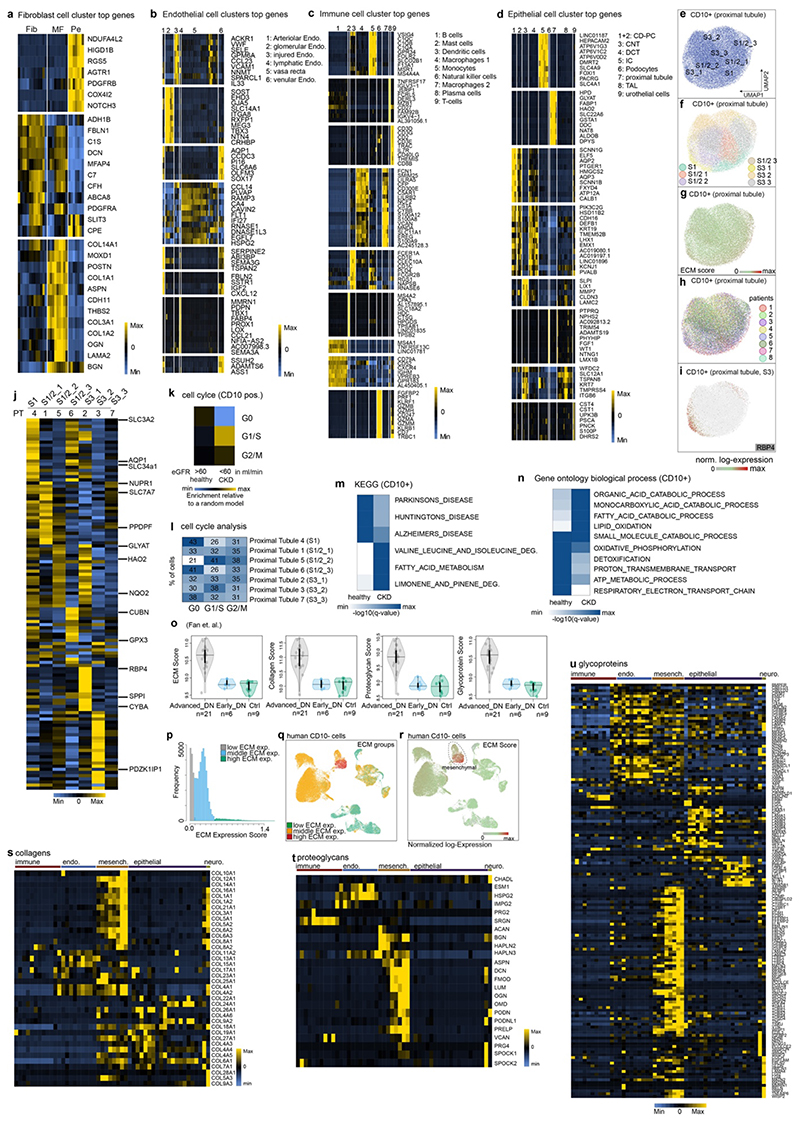
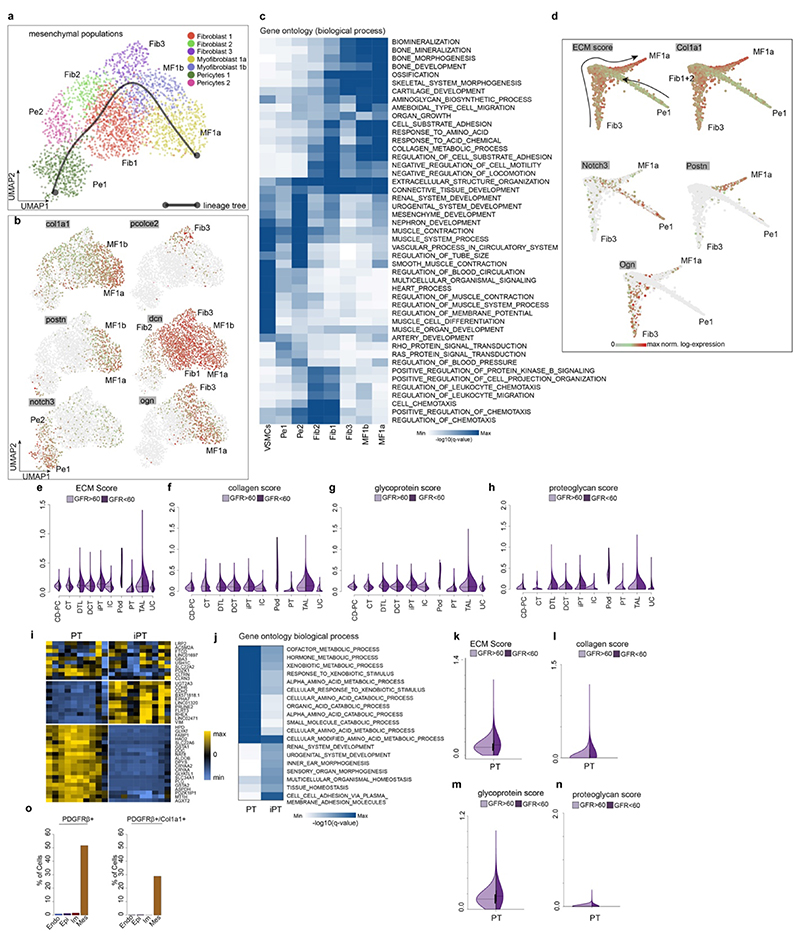
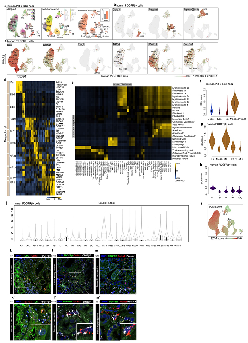
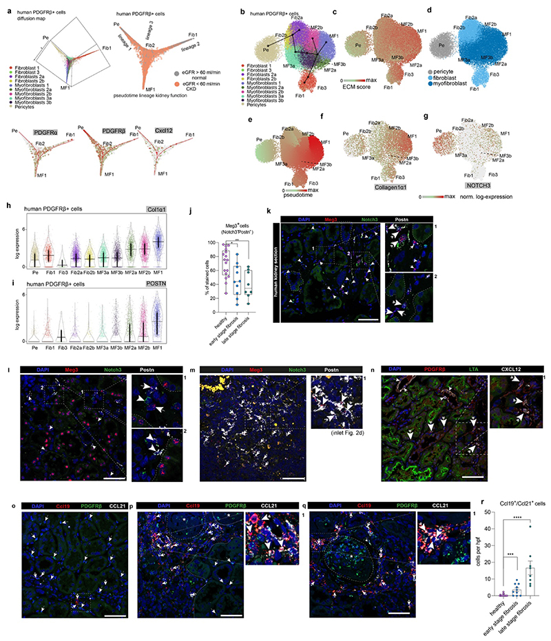
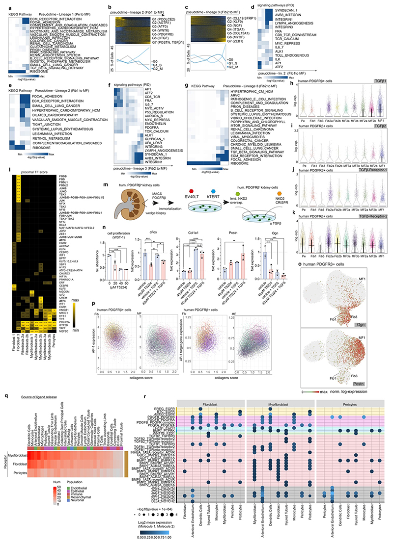
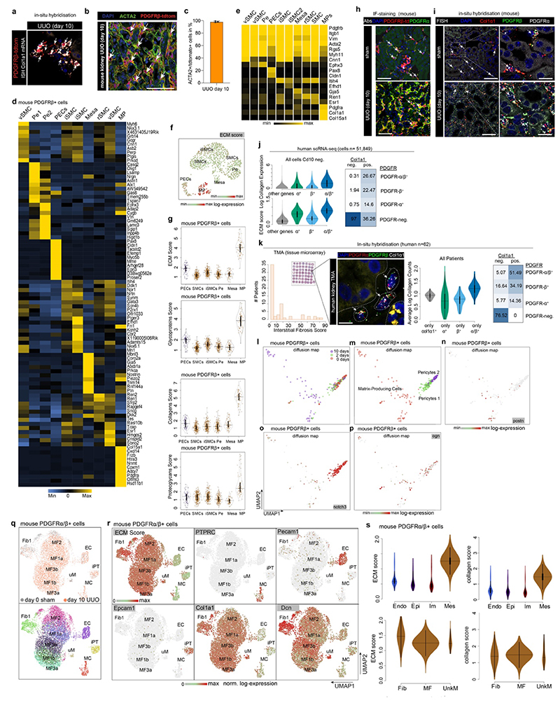
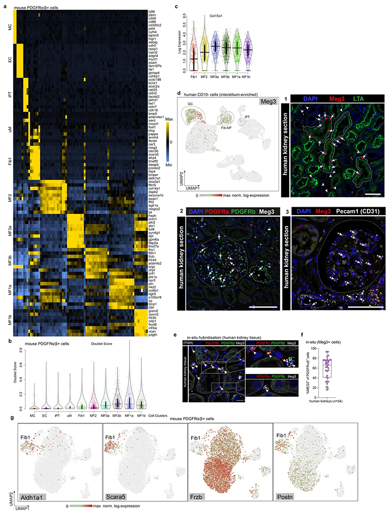
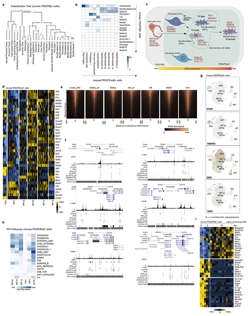
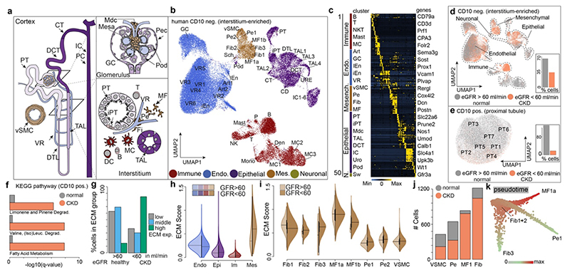
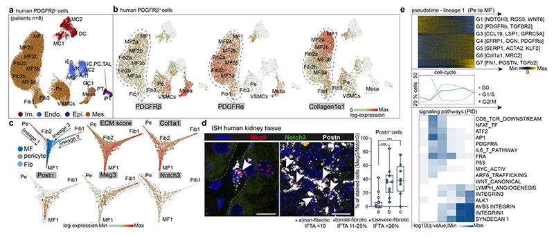
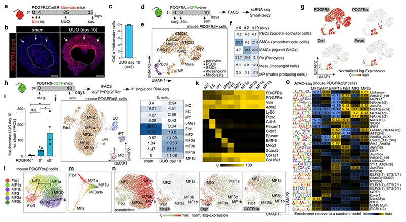
Kidney fibrosis is the hallmark of chronic kidney disease progression; however, at present no antifibrotic therapies exist. The origin, functional heterogeneity and regulation of scar-forming cells that occur during human kidney fibrosis remain poorly understood. Here, using single-cell RNA sequencing, we profiled the transcriptomes of cells from the proximal and non-proximal tubules of healthy and fibrotic human kidneys to map the entire human kidney. This analysis enabled us to map all matrix-producing cells at high resolution, and to identify distinct subpopulations of pericytes and fibroblasts as the main cellular sources of scar-forming myofibroblasts during human kidney fibrosis. We used genetic fate-tracing, time-course single-cell RNA sequencing and ATAC-seq (assay for transposase-accessible chromatin using sequencing) experiments in mice, and spatial transcriptomics in human kidney fibrosis, to shed light on the cellular origins and differentiation of human kidney myofibroblasts and their precursors at high resolution. Finally, we used this strategy to detect potential therapeutic targets, and identified NKD2 as a myofibroblast-specific target in human kidney fibrosis.

摘要

肾纤维化是慢性肾脏病进展的标志;然而,目前尚无抗纤维化疗法。在人类肾纤维化过程中,瘢痕形成细胞的起源、功能异质性和调控仍知之甚少。在这里,我们使用单细胞 RNA 测序技术,对健康和纤维化人类肾脏近端和非近端小管细胞的转录组进行了分析,以绘制整个人类肾脏图谱。这项分析使我们能够以高分辨率绘制所有产生基质的细胞图谱,并鉴定出不同的周细胞和成纤维细胞亚群,它们是人类肾纤维化过程中瘢痕形成肌成纤维细胞的主要细胞来源。我们使用遗传命运追踪、单细胞 RNA 测序时间进程实验和 ATAC-seq(使用测序进行转座酶可及染色质分析)实验在小鼠中进行研究,并在人类肾纤维化中进行空间转录组学研究,以高分辨率揭示人类肾肌成纤维细胞及其前体细胞的细胞起源和分化。最后,我们使用这种策略来检测潜在的治疗靶点,并确定 NKD2 是人类肾纤维化中肌成纤维细胞特异性的靶点。

https://cdn.ncbi.nlm.nih.gov/pmc/blobs/5043/7611626/e62c8b8b0334/EMS114558-f004.jpg
https://cdn.ncbi.nlm.nih.gov/pmc/blobs/5043/7611626/dc5222988248/EMS114558-f005.jpg
https://cdn.ncbi.nlm.nih.gov/pmc/blobs/5043/7611626/42b2948001d1/EMS114558-f006.jpg
https://cdn.ncbi.nlm.nih.gov/pmc/blobs/5043/7611626/aeef8aeaf3fd/EMS114558-f007.jpg
https://cdn.ncbi.nlm.nih.gov/pmc/blobs/5043/7611626/a05464ded1e2/EMS114558-f008.jpg
https://cdn.ncbi.nlm.nih.gov/pmc/blobs/5043/7611626/65ea9aa9335f/EMS114558-f009.jpg
https://cdn.ncbi.nlm.nih.gov/pmc/blobs/5043/7611626/1d67c9073e05/EMS114558-f010.jpg
https://cdn.ncbi.nlm.nih.gov/pmc/blobs/5043/7611626/f696b99835ae/EMS114558-f011.jpg
https://cdn.ncbi.nlm.nih.gov/pmc/blobs/5043/7611626/2885cec842f2/EMS114558-f012.jpg
https://cdn.ncbi.nlm.nih.gov/pmc/blobs/5043/7611626/91b968251579/EMS114558-f013.jpg
https://cdn.ncbi.nlm.nih.gov/pmc/blobs/5043/7611626/00eb38bd5291/EMS114558-f014.jpg
https://cdn.ncbi.nlm.nih.gov/pmc/blobs/5043/7611626/17352a10f927/EMS114558-f001.jpg
https://cdn.ncbi.nlm.nih.gov/pmc/blobs/5043/7611626/0b56e2ed9d3c/EMS114558-f002.jpg
https://cdn.ncbi.nlm.nih.gov/pmc/blobs/5043/7611626/fcfc3c61b9c2/EMS114558-f003.jpg
https://cdn.ncbi.nlm.nih.gov/pmc/blobs/5043/7611626/e62c8b8b0334/EMS114558-f004.jpg

相似文献

1
Decoding myofibroblast origins in human kidney fibrosis.解析人肾纤维化中肌成纤维细胞的起源。
Nature. 2021 Jan;589(7841):281-286. doi: 10.1038/s41586-020-2941-1. Epub 2020 Nov 11.
2
Fate tracing reveals the pericyte and not epithelial origin of myofibroblasts in kidney fibrosis.命运追踪揭示了肾脏纤维化中肌成纤维细胞的周细胞而非上皮细胞起源。
Am J Pathol. 2010 Jan;176(1):85-97. doi: 10.2353/ajpath.2010.090517. Epub 2009 Dec 11.
3
Origin of myofibroblasts and cellular events triggering fibrosis.肌成纤维细胞的起源和触发纤维化的细胞事件。
Kidney Int. 2015 Feb;87(2):297-307. doi: 10.1038/ki.2014.287. Epub 2014 Aug 27.
4
Characterization of human PDGFR-β-positive pericytes from IPF and non-IPF lungs.从特发性肺纤维化和非特发性肺纤维化肺中鉴定人 PDGFR-β 阳性周细胞。
Am J Physiol Lung Cell Mol Physiol. 2018 Dec 1;315(6):L991-L1002. doi: 10.1152/ajplung.00289.2018. Epub 2018 Oct 18.
5
Host responses in tissue repair and fibrosis.组织修复和纤维化中的宿主反应。
Annu Rev Pathol. 2013 Jan 24;8:241-76. doi: 10.1146/annurev-pathol-020712-163930. Epub 2012 Oct 22.
6
Understanding the origin, activation and regulation of matrix-producing myofibroblasts for treatment of fibrotic disease.理解产生细胞外基质的肌成纤维细胞的起源、激活和调控,以治疗纤维化疾病。
J Pathol. 2013 Nov;231(3):273-89. doi: 10.1002/path.4253.
7
Single-cell analysis reveals fibroblast heterogeneity and myofibroblasts in systemic sclerosis-associated interstitial lung disease.单细胞分析揭示了系统性硬皮病相关间质性肺病中成纤维细胞的异质性和肌成纤维细胞。
Ann Rheum Dis. 2019 Oct;78(10):1379-1387. doi: 10.1136/annrheumdis-2018-214865. Epub 2019 Aug 12.
8
Cellular mechanisms of tissue fibrosis. 3. Novel mechanisms of kidney fibrosis.细胞组织纤维化的机制。3. 肾脏纤维化的新机制。
Am J Physiol Cell Physiol. 2013 Apr 1;304(7):C591-603. doi: 10.1152/ajpcell.00414.2012. Epub 2013 Jan 16.
9
[Identification of Biomarkers for Tubular Injury and Interstitial Fibrosis in Chronic Kidney Disease].[慢性肾脏病肾小管损伤和间质纤维化生物标志物的鉴定]
Yakugaku Zasshi. 2017;137(11):1355-1360. doi: 10.1248/yakushi.17-00150.
10
New tools to study renal fibrogenesis.研究肾纤维化的新工具。
Curr Opin Nephrol Hypertens. 2024 Jul 1;33(4):420-426. doi: 10.1097/MNH.0000000000000988. Epub 2024 Apr 8.

引用本文的文献

1
Depletion of tropomyosin 1.6 alleviates TGF-β1-induced fibroblast activation by promoting the α-SMA-MMP9 matrix-degrading structure.原肌球蛋白1.6的缺失通过促进α-平滑肌肌动蛋白-基质金属蛋白酶9基质降解结构来减轻转化生长因子-β1诱导的成纤维细胞活化。
iScience. 2025 Aug 6;28(9):113317. doi: 10.1016/j.isci.2025.113317. eCollection 2025 Sep 19.
2
Regenerative Versus Nonregenerative Repair of the Renal Medulla After Reversal of Ureteral Obstruction.输尿管梗阻解除后肾髓质的再生性修复与非再生性修复
Semin Nephrol. 2025 Aug 23:151669. doi: 10.1016/j.semnephrol.2025.151669.
3
Single-Cell RNA Sequencing Reveals Pericytes Acquire a Fibrotic Phenotype and Promote Mesenteric Adipose Tissue Fibrosis in Crohn's Disease.

本文引用的文献

1
Single-cell RNA-seq reveals ectopic and aberrant lung-resident cell populations in idiopathic pulmonary fibrosis.单细胞 RNA 测序揭示特发性肺纤维化中异位和异常的肺驻留细胞群体。
Sci Adv. 2020 Jul 8;6(28):eaba1983. doi: 10.1126/sciadv.aba1983. eCollection 2020 Jul.
2
Collagen-producing lung cell atlas identifies multiple subsets with distinct localization and relevance to fibrosis.胶原产生肺细胞图谱确定多个具有不同定位和纤维化相关性的亚群。
Nat Commun. 2020 Apr 21;11(1):1920. doi: 10.1038/s41467-020-15647-5.
3
CellPhoneDB: inferring cell-cell communication from combined expression of multi-subunit ligand-receptor complexes.
单细胞RNA测序揭示周细胞获得纤维化表型并促进克罗恩病患者肠系膜脂肪组织纤维化
J Inflamm Res. 2025 Aug 14;18:11055-11071. doi: 10.2147/JIR.S528171. eCollection 2025.
4
Multimodal spatial transcriptomic characterization of mouse kidney injury and repair.小鼠肾损伤与修复的多模态空间转录组学特征分析
Nat Commun. 2025 Aug 14;16(1):7567. doi: 10.1038/s41467-025-62599-9.
5
Fabry Disease Beyond Storage: The Role of Inflammation in Disease Progression.法布里病:超越贮积现象——炎症在疾病进展中的作用
Int J Mol Sci. 2025 Jul 22;26(15):7054. doi: 10.3390/ijms26157054.
6
Analysis of individual patient pathway coordination in a cross-species single-cell kidney atlas.跨物种单细胞肾脏图谱中个体患者路径协调分析
Nat Genet. 2025 Aug 7. doi: 10.1038/s41588-025-02285-0.
7
Inhibitory effects of herbal monomers on ferroptosis in renal fibrosis: a review and mechanistic study.草药单体对肾纤维化中铁死亡的抑制作用:综述与机制研究
Front Pharmacol. 2025 Jul 22;16:1610573. doi: 10.3389/fphar.2025.1610573. eCollection 2025.
8
Complement Proteins Identify Rapidly Progressive Diabetic Kidney Disease.补体蛋白可识别快速进展性糖尿病肾病。
Kidney Int Rep. 2025 May 6;10(7):2296-2310. doi: 10.1016/j.ekir.2025.04.061. eCollection 2025 Jul.
9
Single-cell spatial transcriptomics reveals pathogenic mechanism of renal fibrosis in imiquimod-induced lupus nephritis in mice.单细胞空间转录组学揭示咪喹莫特诱导的小鼠狼疮性肾炎肾纤维化的致病机制。
Biochem Biophys Rep. 2025 Jun 23;43:102087. doi: 10.1016/j.bbrep.2025.102087. eCollection 2025 Sep.
10
Comprehensive analysis of pyroptosis-related gene signatures in renal fibrosis.肾纤维化中焦亡相关基因特征的综合分析
Clin Exp Nephrol. 2025 Jul 3. doi: 10.1007/s10157-025-02726-4.
CellPhoneDB:从多亚基配体-受体复合物的综合表达推断细胞间通讯。
Nat Protoc. 2020 Apr;15(4):1484-1506. doi: 10.1038/s41596-020-0292-x. Epub 2020 Feb 26.
4
Robustness and applicability of transcription factor and pathway analysis tools on single-cell RNA-seq data.转录因子和途径分析工具在单细胞 RNA-seq 数据上的稳健性和适用性。
Genome Biol. 2020 Feb 12;21(1):36. doi: 10.1186/s13059-020-1949-z.
5
Resolving the fibrotic niche of human liver cirrhosis at single-cell level.解析人肝硬化的纤维性龛位于单细胞水平。
Nature. 2019 Nov;575(7783):512-518. doi: 10.1038/s41586-019-1631-3. Epub 2019 Oct 9.
6
Comparison of Kidney Transcriptomic Profiles of Early and Advanced Diabetic Nephropathy Reveals Potential New Mechanisms for Disease Progression.早期和晚期糖尿病肾病的肾脏转录组谱比较揭示了疾病进展的潜在新机制。
Diabetes. 2019 Dec;68(12):2301-2314. doi: 10.2337/db19-0204. Epub 2019 Oct 2.
7
Benchmark and integration of resources for the estimation of human transcription factor activities.用于估计人类转录因子活性的资源的基准测试和整合。
Genome Res. 2019 Aug;29(8):1363-1375. doi: 10.1101/gr.240663.118. Epub 2019 Jul 24.
8
A single-nucleus RNA-sequencing pipeline to decipher the molecular anatomy and pathophysiology of human kidneys.一种用于解析人类肾脏分子解剖结构和病理生理学的单细胞核 RNA 测序工作流程。
Nat Commun. 2019 Jun 27;10(1):2832. doi: 10.1038/s41467-019-10861-2.
9
CHETAH: a selective, hierarchical cell type identification method for single-cell RNA sequencing.CHETAH:一种用于单细胞 RNA 测序的选择性、层次化细胞类型识别方法。
Nucleic Acids Res. 2019 Sep 19;47(16):e95. doi: 10.1093/nar/gkz543.
10
Comprehensive Integration of Single-Cell Data.单细胞数据的综合整合。
Cell. 2019 Jun 13;177(7):1888-1902.e21. doi: 10.1016/j.cell.2019.05.031. Epub 2019 Jun 6.