Department of Internal Medicine, University of Texas Medical Branch, Galveston, Texas, United States of America.
Department of Microbiology and Immunology, University of Texas Medical Branch, Galveston, Texas, United States of America.
PLoS One. 2020 Nov 12;15(11):e0242337. doi: 10.1371/journal.pone.0242337. eCollection 2020.
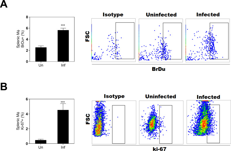
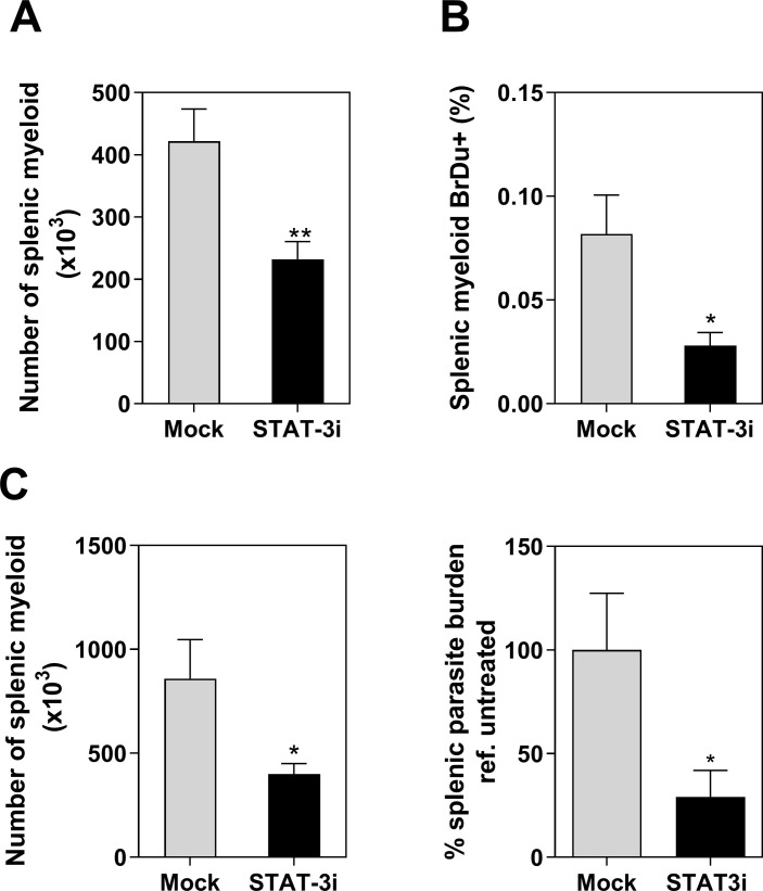
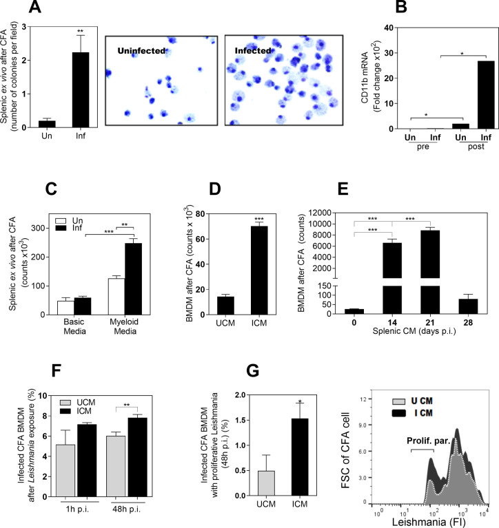
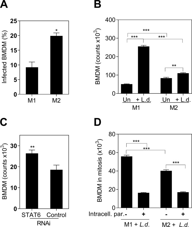
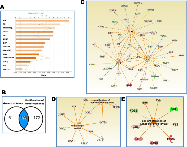
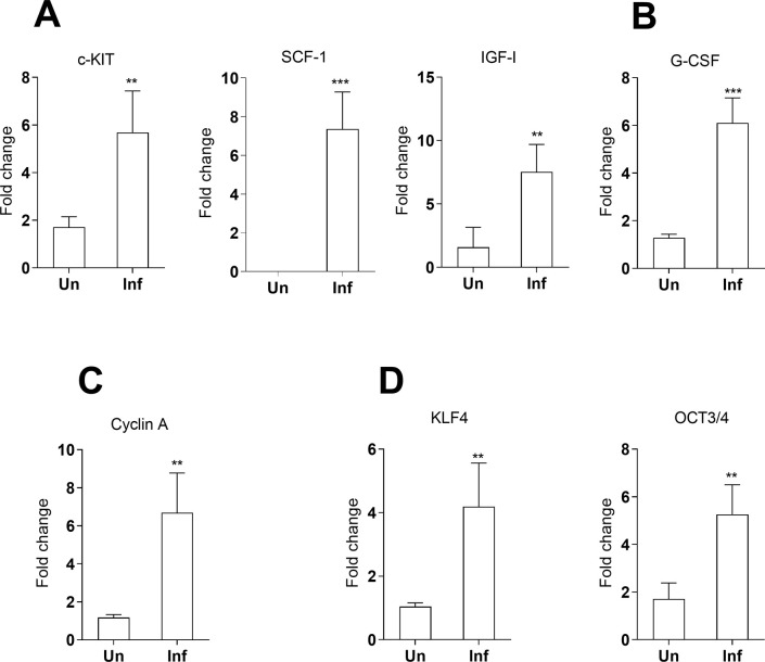
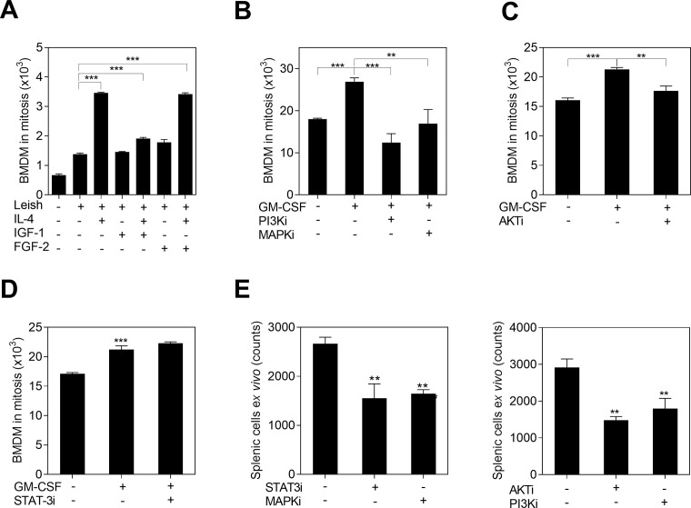
Visceral leishmaniasis (VL) is characterized by expansion of myeloid cells in the liver and spleen, which leads to a severe splenomegaly associated with higher risk of mortality. This increased cellularity is thought to be a consequence of recruitment of cells to the viscera. We studied whether the local proliferation of splenic myeloid cells contributes to increased splenic cellularity. We found that a monocyte-like population of adherent splenic cells from Leishmania donovani-infected hamsters had enhanced replicative capacity ex vivo and in vivo (BrdU incorporation, p<0.0001). In vitro assays demonstrated that proliferation was more pronounced in the proinflammatory M1 environment and that intracellular infection prevented proliferation. Secondary analysis of the published splenic transcriptome in the hamster model of progressive VL revealed a gene expression signature that included division of tumoral cells (Z = 2.0), cell cycle progression (Z = 2.3), hematopoiesis (Z = 2.8), proliferation of stem cells (Z = 2.5) and overexpression of proto-oncogenes. Regulators of myeloid cell proliferation were predicted in-silico (CSF2, TLR4, IFNG, IL-6, IL-4, RTK signaling, and STAT3). The in-silico prediction was confirmed with chemical inhibitors of PI3K/AKT, MAPK and STAT3 which decreased splenic myeloid cell division ex vivo. Hamsters infected with L. donovani treated with a STAT3 inhibitor had reduced in situ splenic myeloid proliferation (p = 0.03) and parasite burden. We conclude that monocyte-like myeloid cells have increased STAT3-dependent proliferation in the spleen of hamsters with visceral leishmaniasis and that inhibition of STAT3 reduces myeloid cell proliferation and parasite burden.
内脏利什曼病(VL)的特征是肝脏和脾脏中髓样细胞的扩张,导致严重的脾肿大,与更高的死亡率相关。这种细胞增多被认为是细胞向内脏招募的结果。我们研究了脾脏髓样细胞的局部增殖是否有助于增加脾脏细胞的数量。我们发现,来自感染杜氏利什曼原虫的仓鼠的贴壁脾脏细胞中的单核细胞样群体具有增强的体外和体内复制能力(BrdU 掺入,p<0.0001)。体外试验表明,在促炎 M1 环境中增殖更为明显,并且细胞内感染阻止了增殖。对仓鼠进展性 VL 模型中已发表的脾脏转录组的二次分析显示,包含肿瘤细胞分裂(Z = 2.0)、细胞周期进程(Z = 2.3)、造血(Z = 2.8)、干细胞增殖(Z = 2.5)和原癌基因过表达的基因表达特征。预测了骨髓细胞增殖的调节剂(CSF2、TLR4、IFNG、IL-6、IL-4、RTK 信号和 STAT3)。通过化学抑制剂 PI3K/AKT、MAPK 和 STAT3 进行的计算机预测证实,这些抑制剂可减少体外脾脏髓样细胞的分裂。用 STAT3 抑制剂治疗的感染杜氏利什曼原虫的仓鼠,其脾脏原位髓样细胞增殖减少(p = 0.03)和寄生虫负担减少。我们得出结论,单核细胞样髓样细胞在患有内脏利什曼病的仓鼠脾脏中具有增加的 STAT3 依赖性增殖,并且抑制 STAT3 可减少髓样细胞增殖和寄生虫负担。