Sun Chang-Yu, Stifler Cayla A, Chopdekar Rajesh V, Schmidt Connor A, Parida Ganesh, Schoeppler Vanessa, Fordyce Benjamin I, Brau Jack H, Mass Tali, Tambutté Sylvie, Gilbert Pupa U P A
Department of Physics, University of Wisconsin, Madison, WI 53706.
Advanced Light Source, Lawrence Berkeley National Laboratory, Berkeley, CA 94720.
Proc Natl Acad Sci U S A. 2020 Dec 1;117(48):30159-30170. doi: 10.1073/pnas.2012025117. Epub 2020 Nov 13.
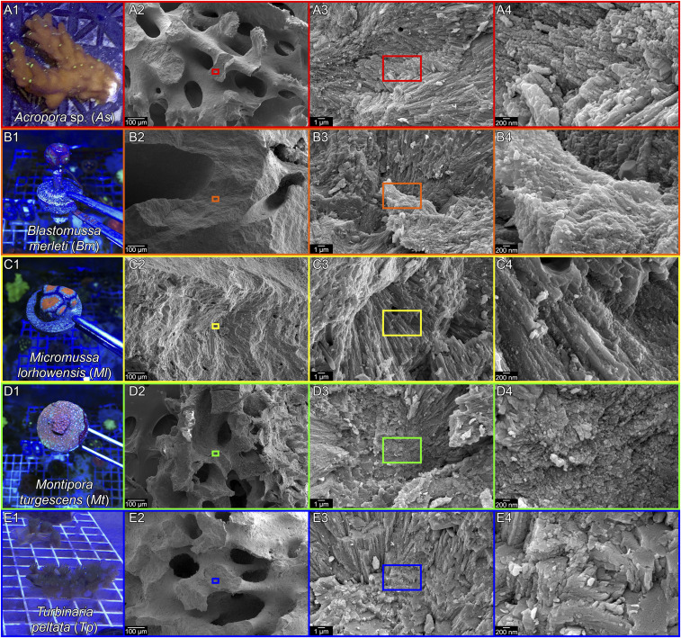
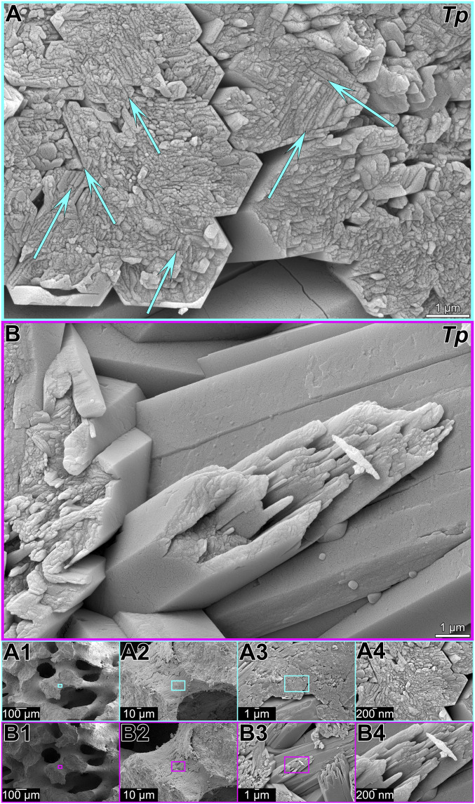
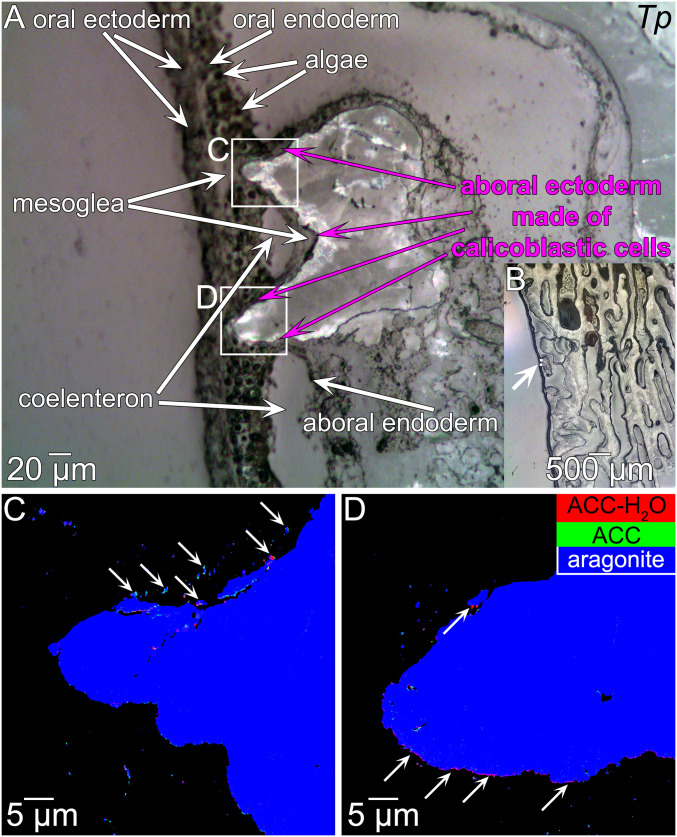
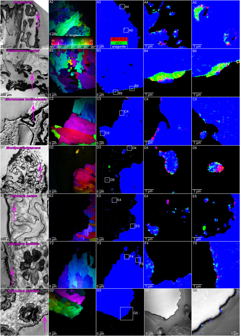
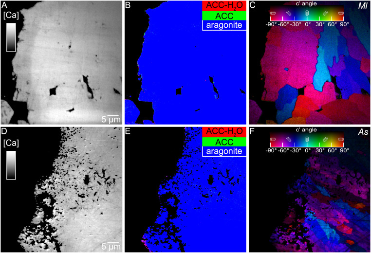
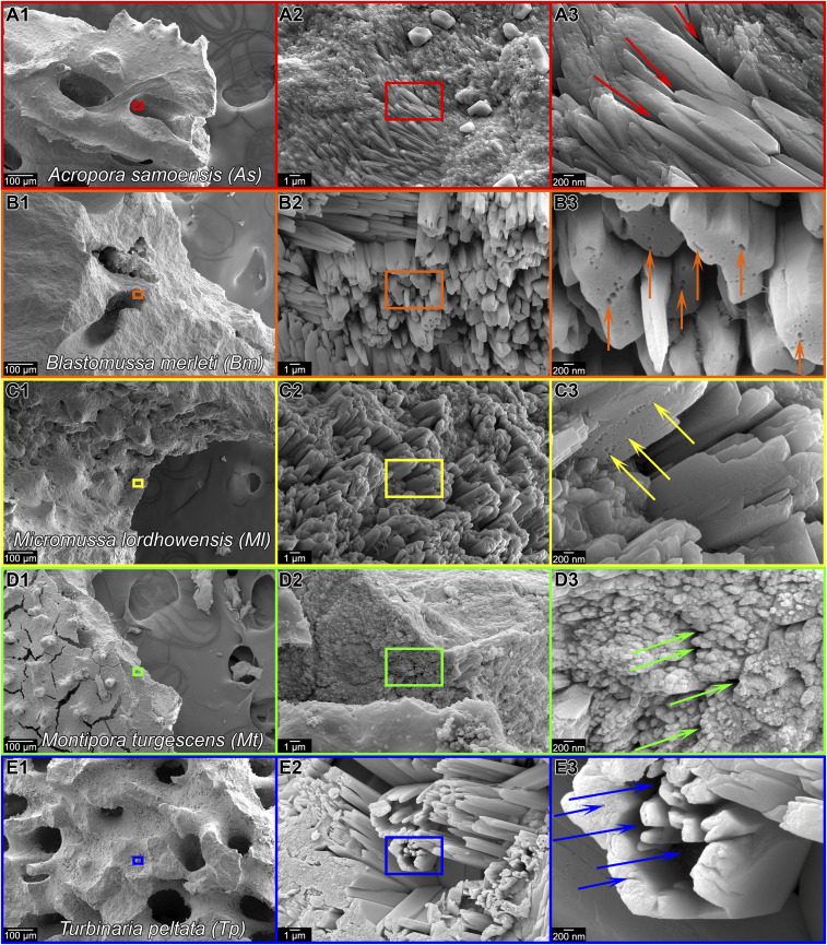
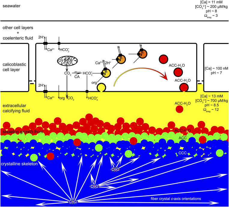
Reef-building corals and their aragonite (CaCO) skeletons support entire reef ecosystems, yet their formation mechanism is poorly understood. Here we used synchrotron spectromicroscopy to observe the nanoscale mineralogy of fresh, forming skeletons from six species spanning all reef-forming coral morphologies: Branching, encrusting, massive, and table. In all species, hydrated and anhydrous amorphous calcium carbonate nanoparticles were precursors for skeletal growth, as previously observed in a single species. The amorphous precursors here were observed in tissue, between tissue and skeleton, and at growth fronts of the skeleton, within a low-density nano- or microporous layer varying in thickness from 7 to 20 µm. Brunauer-Emmett-Teller measurements, however, indicated that the mature skeletons at the microscale were space-filling, comparable to single crystals of geologic aragonite. Nanoparticles alone can never fill space completely, thus ion-by-ion filling must be invoked to fill interstitial pores. Such ion-by-ion diffusion and attachment may occur from the supersaturated calcifying fluid known to exist in corals, or from a dense liquid precursor, observed in synthetic systems but never in biogenic ones. Concomitant particle attachment and ion-by-ion filling was previously observed in synthetic calcite rhombohedra, but never in aragonite pseudohexagonal prisms, synthetic or biogenic, as observed here. Models for biomineral growth, isotope incorporation, and coral skeletons' resilience to ocean warming and acidification must take into account the dual formation mechanism, including particle attachment and ion-by-ion space filling.
造礁珊瑚及其文石(CaCO)骨骼支撑着整个珊瑚礁生态系统,但其形成机制却鲜为人知。在这里,我们使用同步加速器光谱显微镜观察了六种具有所有造礁珊瑚形态的新鲜、正在形成的骨骼的纳米级矿物学特征:分支状、覆盖状、块状和板状。在所有物种中,水合和无水无定形碳酸钙纳米颗粒都是骨骼生长的前体,这与之前在单一物种中观察到的情况一致。这里观察到无定形前体存在于组织中、组织与骨骼之间以及骨骼的生长前沿,位于一个厚度从7到20微米不等的低密度纳米或微孔层内。然而,布鲁诺尔-埃米特-泰勒测量表明,微观尺度上的成熟骨骼是充满空间的,类似于地质文石的单晶。仅纳米颗粒永远无法完全填满空间,因此必须通过逐个离子填充来填充间隙孔隙。这种逐个离子的扩散和附着可能发生在已知存在于珊瑚中的过饱和钙化液中,或者发生在合成系统中观察到但在生物系统中从未观察到的致密液体前体中。之前在合成方解石菱面体中观察到了伴随颗粒附着和逐个离子填充的情况,但在这里观察到,无论是合成的还是生物成因的文石假六方棱柱中都从未观察到这种情况。生物矿化生长、同位素掺入以及珊瑚骨骼对海洋变暖和酸化的恢复力模型必须考虑到这种双重形成机制,包括颗粒附着和逐个离子的空间填充。