From the Smidt Heart Institute, Cedars-Sinai Medical Center, 127 S San Vicente Blvd, AHSP, Suite A3600, Los Angeles, CA 90048-0750 (A.C.K.); Department of Radiology and Imaging Sciences, Emory University, Atlanta, Ga (A.P.); Winship Cancer Institute, Emory University, Atlanta, Ga (A.P.); Department of Biomedical Engineering, Georgia Institute of Technology-Emory University, Atlanta, Ga (A.P.); Department of Physics and Astronomy, Johns Hopkins University, Baltimore, Md (D.S.); Extreme Light Infrastructure-Nuclear Physics, Bucharest-Magurele, Romania (D.S.); Department of Radiology, University of Wisconsin School of Medicine and Public Health, Madison, Wis (D.A.B.); and Department of Cardiology, The Johns Hopkins Hospital, Baltimore, Md (J.A.C.L.).
Radiology. 2021 Jan;298(1):3-17. doi: 10.1148/radiol.2020192791. Epub 2020 Nov 17.
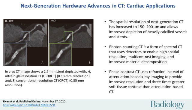
Impending major hardware advances in cardiac CT include three areas: ultra-high-resolution (UHR) CT, photon-counting CT, and phase-contrast CT. Cardiac CT is a particularly demanding CT application that requires a high degree of temporal resolution, spatial resolution, and soft-tissue contrast in a moving structure. In this review, cardiac CT is used to highlight the strengths of these technical advances. UHR CT improves visualization of calcified and stented vessels but may result in increased noise and radiation exposure. Photon-counting CT uses multiple photon energies to reduce artifacts, improve contrast resolution, and perform material decomposition. Finally, phase-contrast CT uses x-ray refraction properties to improve spatial and soft-tissue contrast. This review describes these hardware advances in CT and their relevance to cardiovascular imaging.
心脏 CT 即将迎来的主要硬件进展包括三个方面:超高分辨率(UHR)CT、光子计数 CT 和相位对比 CT。心脏 CT 是一种特别苛刻的 CT 应用,需要在运动结构中具有高度的时间分辨率、空间分辨率和软组织对比度。在本综述中,心脏 CT 被用于突出这些技术进步的优势。UHR CT 改善了钙化和支架血管的可视化效果,但可能导致噪声增加和辐射暴露增加。光子计数 CT 使用多个光子能量来减少伪影、提高对比分辨率和进行材料分解。最后,相位对比 CT 使用 X 射线折射特性来提高空间和软组织对比度。本综述描述了 CT 中的这些硬件进展及其与心血管成像的相关性。