Cameli Matteo, Miglioranza Marcelo Haertel, Magne Julien, Mandoli Giulia Elena, Benfari Giovanni, Ancona Roberta, Sibilio Gerolamo, Reskovic Luksic Vlatka, Dejan Dosen, Griseli Leonardo, Van De Heyning Caroline M, Mortelmans Philippe, Michalski Blazej, Kupczynska Karolina, Di Giannuario Giovanna, Devito Fiorella, Dulgheru Raluca, Ilardi Federica, Salustri Alessandro, Abushahba Galal, Morrone Doralisa, Fabiani Iacopo, Penicka Martin, Katbeh Asim, Sammarco Giuseppe, Esposito Roberta, Santoro Ciro, Pastore Maria Concetta, Comenale Pinto Salvatore, Kalinin Artem, Pičkure Žanna, Ažman Juvan Katja, Zupan Mežnar Anja, Coisne Augustine, Coppin Amandine, Opris Mihaela Maria, Nistor Dan Octavian, Paakkanen Riitta, Biering-Sørensen Tor, Olsen Flemming Javier, Lapinskas Tomas, Vaškelyté Jolanta Justina, Galian-Gay Laura, Casas Guillem, Motoc Andreea Iulia, Papadopoulos Constantinos Hristou, Loizos Savvas, Ágoston Gergely, Szabó Istvan, Hristova Krasimira, Tsonev Svetlin Netkov, Galli Elena, Vinereanu Dragos, Mihaila Baldea Sorina, Muraru Denisa, Mondillo Sergio, Donal Erwan, Galderisi Maurizio, Cosyns Bernard, Edvardsen Thor, Popescu Bogdan A
Department of Medical Biotechnologies, Division of Cardiology, University of Siena, 53100 Siena, Italy.
Institute of Cardiology, University Foundation of Cardiology, Porto Alegre 90620-000, Brazil.
Diagnostics (Basel). 2020 Nov 13;10(11):946. doi: 10.3390/diagnostics10110946.
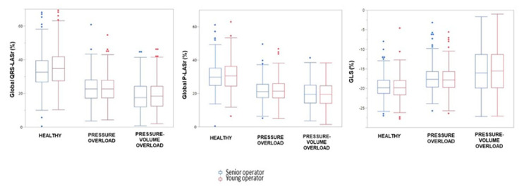
Two methods are currently available for left atrial (LA) strain measurement by speckle tracking echocardiography, with two different reference timings for starting the analysis: QRS (QRS-LASr) and P wave (P-LASr). The aim of MASCOT HIT study was to define which of the two was more reproducible, more feasible, and less time consuming. In 26 expert centers, LA strain was analyzed by two different echocardiographers (young vs senior) in a blinded fashion. The study population included: healthy subjects, patients with arterial hypertension or aortic stenosis (LA pressure overload, group 2) and patients with mitral regurgitation or heart failure (LA volume-pressure overload, group 3). Difference between the inter-correlation coefficient (ICC) by the two echocardiographers using the two techniques, feasibility and analysis time of both methods were analyzed. A total of 938 subjects were included: 309 controls, 333 patients in group 2, and 296 patients in group 3. The ICC was comparable between QRS-LASr (0.93) and P-LASr (0.90). The young echocardiographers calculated QRS-LASr in 90% of cases, the expert ones in 95%. The feasibility of P-LASr was 85% by young echocardiographers and 88% by senior ones. QRS-LASr young median time was 110 s (interquartile range, IR, 78-149) vs senior 110 s (IR 78-155); for P-LASr, 120 s (IR 80-165) and 120 s (IR 90-161), respectively. LA strain was feasible in the majority of patients with similar reproducibility for both methods. QRS complex guaranteed a slightly higher feasibility and a lower time wasting compared to the use of P wave as the reference.
目前,通过斑点追踪超声心动图测量左心房(LA)应变有两种方法,且起始分析的参考时间不同:QRS波(QRS-LASr)和P波(P-LASr)。MASCOT HIT研究的目的是确定这两种方法中哪种更具可重复性、更可行且耗时更少。在26个专家中心,由两名不同的超声心动图检查人员(年轻与资深)以盲法分析LA应变。研究人群包括:健康受试者、动脉高血压或主动脉瓣狭窄患者(LA压力过载,第2组)以及二尖瓣反流或心力衰竭患者(LA容量-压力过载,第3组)。分析了两名超声心动图检查人员使用这两种技术时的组内相关系数(ICC)差异、两种方法的可行性和分析时间。总共纳入了938名受试者:309名对照组、第2组的333名患者和第3组的296名患者。QRS-LASr(0.93)和P-LASr(0.90)的ICC相当。年轻的超声心动图检查人员在90%的病例中计算QRS-LASr,专家在95%的病例中计算。年轻的超声心动图检查人员对P-LASr的可行性为85%,资深人员为88%。QRS-LASr年轻人员的中位时间为110秒(四分位间距,IR,78 - 149),资深人员为110秒(IR 78 - 155);对于P-LASr,分别为120秒(IR 80 - 165)和120秒(IR 90 - 161)。LA应变在大多数患者中是可行的,两种方法的可重复性相似。与以P波作为参考相比,QRS波保证了略高的可行性和更低的时间消耗。