• 文献检索
  • 文档翻译
  • 深度研究
  • 学术资讯
  • Suppr Zotero 插件Zotero 插件
  • 邀请有礼
  • 套餐&价格
  • 历史记录
应用&插件
Suppr Zotero 插件Zotero 插件浏览器插件Mac 客户端Windows 客户端微信小程序
定价
高级版会员购买积分包购买API积分包
服务
文献检索文档翻译深度研究API 文档MCP 服务
关于我们
关于 Suppr公司介绍联系我们用户协议隐私条款
关注我们

Suppr 超能文献

核心技术专利:CN118964589B侵权必究
粤ICP备2023148730 号-1Suppr @ 2026

文献检索

告别复杂PubMed语法,用中文像聊天一样搜索,搜遍4000万医学文献。AI智能推荐,让科研检索更轻松。

立即免费搜索

文件翻译

保留排版,准确专业,支持PDF/Word/PPT等文件格式,支持 12+语言互译。

免费翻译文档

深度研究

AI帮你快速写综述,25分钟生成高质量综述,智能提取关键信息,辅助科研写作。

立即免费体验

轻度阿尔茨海默病患者的眼部血管变化:中心凹无血管区、脉络膜厚度及视盘血红蛋白分析

Ocular Vascular Changes in Mild Alzheimer's Disease Patients: Foveal Avascular Zone, Choroidal Thickness, and ONH Hemoglobin Analysis.

作者信息

Salobrar-Garcia Elena, Méndez-Hernández Carmen, Hoz Rosa de, Ramírez Ana I, López-Cuenca Inés, Fernández-Albarral José A, Rojas Pilar, Wang Surina, García-Feijoo Julián, Gil Pedro, Salazar Juan J, Ramírez José M

机构信息

Instituto de Investigaciones Oftalmológicas Ramón Castroviejo, Universidad Complutense de Madrid (UCM), 28040 Madrid, Spain.

Facultad de Óptica y Optometría, Departamento de Inmunología, Oftalmología y ORL, UCM, 28037 Madrid, Spain.

出版信息

J Pers Med. 2020 Nov 15;10(4):231. doi: 10.3390/jpm10040231.

DOI:10.3390/jpm10040231
PMID:33203157
原文链接:https://pmc.ncbi.nlm.nih.gov/articles/PMC7712569/
Abstract

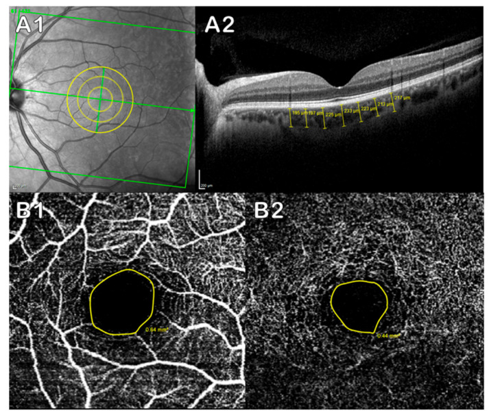
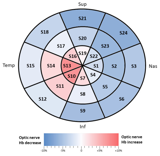
In Alzheimer's disease (AD), vascular changes could be caused by amyloid beta (Aβ) aggregates replacing the contractile smooth musculature of the arteriole walls. These changes happen in the brain vascular network, but also in the eye, and are related to decreased vascular density and low blood flow. In patients with Alzheimer's disease, thinning of the choroid and the retina has been shown. The aim of this prospective study was to assess the retinal and choroidal vascular systems, analyzing the choroidal thickness with optical coherence tomography (OCT), the foveal avascular zone (FAZ) with OCT-angiography (OCTA), and the optic nerve head (ONH) hemoglobin with the Laguna ONhE program, to evaluate which of the two ocular vascular systems shows earlier changes in mild AD patients. These patients, compared to controls, showed a significantly thinner choroid at all the analyzed points, with the exception of the temporal macula (at 1000 and 1500 µm from the fovea). On the other hand, the FAZ and ONH hemoglobin did not show significant differences. In conclusion, a thinner choroid was the main ocular vascular change observed in mild AD patients, while the retinal vessels were not yet affected. Therefore, choroidal thickness could be used an early biomarker in AD.

摘要

在阿尔茨海默病(AD)中,血管变化可能是由淀粉样β蛋白(Aβ)聚集体取代小动脉壁的收缩性平滑肌组织引起的。这些变化不仅发生在脑血管网络中,也发生在眼睛中,并且与血管密度降低和血流减少有关。在阿尔茨海默病患者中,已显示脉络膜和视网膜变薄。这项前瞻性研究的目的是评估视网膜和脉络膜血管系统,通过光学相干断层扫描(OCT)分析脉络膜厚度,通过OCT血管造影(OCTA)分析黄斑无血管区(FAZ),并使用Laguna ONhE程序分析视神经乳头(ONH)血红蛋白,以评估在轻度AD患者中两个眼血管系统中哪个显示出更早的变化。与对照组相比,这些患者在所有分析点的脉络膜均明显变薄,但颞侧黄斑(距黄斑中心凹1000和1500 µm处)除外。另一方面,FAZ和ONH血红蛋白没有显示出显著差异。总之,脉络膜变薄是在轻度AD患者中观察到的主要眼血管变化,而视网膜血管尚未受到影响。因此,脉络膜厚度可作为AD的早期生物标志物。

https://cdn.ncbi.nlm.nih.gov/pmc/blobs/1cc3/7712569/9e9cbd4a1a69/jpm-10-00231-g002.jpg
https://cdn.ncbi.nlm.nih.gov/pmc/blobs/1cc3/7712569/38dac0b60460/jpm-10-00231-g001.jpg
https://cdn.ncbi.nlm.nih.gov/pmc/blobs/1cc3/7712569/9e9cbd4a1a69/jpm-10-00231-g002.jpg
https://cdn.ncbi.nlm.nih.gov/pmc/blobs/1cc3/7712569/38dac0b60460/jpm-10-00231-g001.jpg
https://cdn.ncbi.nlm.nih.gov/pmc/blobs/1cc3/7712569/9e9cbd4a1a69/jpm-10-00231-g002.jpg

相似文献

1
Ocular Vascular Changes in Mild Alzheimer's Disease Patients: Foveal Avascular Zone, Choroidal Thickness, and ONH Hemoglobin Analysis.轻度阿尔茨海默病患者的眼部血管变化:中心凹无血管区、脉络膜厚度及视盘血红蛋白分析
J Pers Med. 2020 Nov 15;10(4):231. doi: 10.3390/jpm10040231.
2
Foveal Avascular Zone and Choroidal Thickness Are Decreased in Subjects with Hard Drusen and without High Genetic Risk of Developing Alzheimer's Disease.在患有硬性玻璃膜疣且无阿尔茨海默病高遗传风险的受试者中,黄斑无血管区和脉络膜厚度降低。
Biomedicines. 2021 Jun 2;9(6):638. doi: 10.3390/biomedicines9060638.
3
Choroidal thinning as a new finding in Alzheimer's disease: evidence from enhanced depth imaging spectral domain optical coherence tomography.脉络膜变薄作为阿尔茨海默病的一项新发现:来自增强深度成像光谱域光学相干断层扫描的证据
J Alzheimers Dis. 2014;40(4):907-17. doi: 10.3233/JAD-132039.
4
Use of optical coherence tomography angiography for assessment of microvascular changes in the macula and optic nerve head in hypertensive patients without hypertensive retinopathy.应用光相干断层扫描血管造影评估无高血压性视网膜病变的高血压患者的黄斑和视神经乳头的微血管变化。
Microvasc Res. 2020 May;129:103969. doi: 10.1016/j.mvr.2019.103969. Epub 2019 Dec 23.
5
Optical coherence tomography of retinal and choroidal layers in patients with familial hypercholesterolaemia treated with lipoprotein apheresis.接受脂蛋白分离治疗的家族性高胆固醇血症患者视网膜和脉络膜层的光学相干断层扫描
Atheroscler Suppl. 2019 Dec;40:49-54. doi: 10.1016/j.atherosclerosissup.2019.08.031.
6
Sex-Related Variations of Retinal and Choroidal Thickness and Foveal Avascular Zone in Healthy and Diabetic Children Assessed by Optical Coherence Tomography Imaging.通过光学相干断层扫描成像评估健康和糖尿病儿童视网膜和脉络膜厚度以及黄斑无血管区的性别差异
Ophthalmologica. 2019;241(3):173-178. doi: 10.1159/000495622. Epub 2019 Jan 16.
7
Retinal vascular density evaluated by optical coherence tomography angiography in macular telangiectasia type 2.通过光学相干断层扫描血管造影术评估2型黄斑毛细血管扩张症的视网膜血管密度。
Int Ophthalmol. 2019 Oct;39(10):2245-2256. doi: 10.1007/s10792-018-01060-x. Epub 2019 Jan 3.
8
Evaluation of optical coherence tomography angiographic findings in Alzheimer's type dementia.阿尔茨海默型痴呆的光学相干断层扫描血管造影结果评估
Br J Ophthalmol. 2018 Feb;102(2):233-237. doi: 10.1136/bjophthalmol-2017-310476. Epub 2017 Jun 9.
9
Optical Coherence Tomography Angiography of Macula and Optic Nerve in Autism Spectrum Disorder: A Pilot Study.自闭症谱系障碍中黄斑和视神经的光学相干断层扫描血管造影:一项初步研究。
J Clin Med. 2020 Sep 27;9(10):3123. doi: 10.3390/jcm9103123.
10
Changes in retina and choroid after haemodialysis assessed using optical coherence tomography angiography.使用光学相干断层扫描血管造影术评估血液透析后视网膜和脉络膜的变化。
Clin Exp Optom. 2018 Sep;101(5):674-679. doi: 10.1111/cxo.12660. Epub 2018 Jan 22.

引用本文的文献

1
Ocular changes as potential biomarkers for early diagnosis of Alzheimer's disease.眼部变化作为阿尔茨海默病早期诊断的潜在生物标志物。
Alzheimers Dement. 2025 Aug;21(8):e70476. doi: 10.1002/alz.70476.
2
Choroidal evaluation of FTLD-Tau and biomarker-determined Alzheimer's disease.额颞叶痴呆- Tau蛋白病和生物标志物确定的阿尔茨海默病的脉络膜评估
Sci Rep. 2025 Jul 1;15(1):22290. doi: 10.1038/s41598-025-02020-z.
3
Retinal vascular biomarkers in mild cognitive impairment and Alzheimer's disease: a comprehensive review and meta-analysis.轻度认知障碍和阿尔茨海默病中的视网膜血管生物标志物:一项全面综述和荟萃分析。

本文引用的文献

1
Spatial analysis of thickness changes in ten retinal layers of Alzheimer's disease patients based on optical coherence tomography.基于光学相干断层扫描的阿尔茨海默病患者 10 层视网膜厚度变化的空间分析。
Sci Rep. 2019 Sep 10;9(1):13000. doi: 10.1038/s41598-019-49353-0.
2
Identification of Retinal Biomarkers in Alzheimer's Disease Using Optical Coherence Tomography: Recent Insights, Challenges, and Opportunities.利用光学相干断层扫描技术识别阿尔茨海默病中的视网膜生物标志物:最新见解、挑战与机遇
J Clin Med. 2019 Jul 9;8(7):996. doi: 10.3390/jcm8070996.
3
Is retinal vasculature a biomarker in amyloid proven Alzheimer's disease?
Alzheimers Dement (Amst). 2025 Jun 16;17(2):e70132. doi: 10.1002/dad2.70132. eCollection 2025 Apr-Jun.
4
Choroidal Vascular Volume, White Matter Hyperintensity, and Their Interaction With Cognitive Function in Aging Adults.脉络膜血管容积、白质高信号及其与老年人认知功能的相互作用
J Am Heart Assoc. 2025 Jun 17;14(12):e039369. doi: 10.1161/JAHA.124.039369. Epub 2025 Jun 11.
5
Retinal microvascular density and inner thickness in Alzheimer's disease and mild cognitive impairment.阿尔茨海默病和轻度认知障碍患者的视网膜微血管密度及内层厚度
Front Aging Neurosci. 2025 Feb 28;17:1477008. doi: 10.3389/fnagi.2025.1477008. eCollection 2025.
6
Optical Coherence Tomography in Huntington's Disease-A Potential Future Biomarker for Neurodegeneration?光学相干断层扫描在亨廷顿舞蹈病中的应用——一种未来潜在的神经退行性变生物标志物?
Neurol Int. 2025 Jan 20;17(1):13. doi: 10.3390/neurolint17010013.
7
Changes in choroidal thickness quantified by Optical Coherence Tomography across cognitive impairment: data from the NORFACE cohort.光学相干断层扫描定量测量脉络膜厚度在认知障碍中的变化:NORFACE 队列研究数据。
Alzheimers Res Ther. 2024 Nov 16;16(1):249. doi: 10.1186/s13195-024-01616-3.
8
Feature-Based Classification of Mild Cognitive Impairment and Alzheimer's Disease Based on Optical Coherence Tomographic Angiographic Image.基于光学相干断层扫描血管造影图像的轻度认知障碍和阿尔茨海默病的特征分类。
Sensors (Basel). 2024 Aug 11;24(16):5192. doi: 10.3390/s24165192.
9
Unlocking the Potential of Vessel Density and the Foveal Avascular Zone in Optical Coherence Tomography Angiography as Biomarkers in Alzheimer's Disease.揭示光学相干断层扫描血管造影中血管密度和黄斑无血管区作为阿尔茨海默病生物标志物的潜力。
Healthcare (Basel). 2024 Aug 9;12(16):1589. doi: 10.3390/healthcare12161589.
10
Retinal Vascular and Structural Changes in the Murine Alzheimer's Model from 6 to 20 Months.从 6 个月到 20 个月的小鼠阿尔茨海默病模型中的视网膜血管和结构变化。
Biomolecules. 2024 Jul 10;14(7):828. doi: 10.3390/biom14070828.
视网膜血管系统是经淀粉样蛋白证实的阿尔茨海默病的生物标志物吗?
Alzheimers Dement (Amst). 2019 May 10;11:383-391. doi: 10.1016/j.dadm.2019.03.006. eCollection 2019 Dec.
4
Optical coherence tomography angiography in preclinical Alzheimer's disease.光学相干断层扫描血管造影在临床前阿尔茨海默病中的应用。
Br J Ophthalmol. 2020 Feb;104(2):157-161. doi: 10.1136/bjophthalmol-2019-314127. Epub 2019 May 22.
5
Functional and morphological changes of the retinal vessels in Alzheimer's disease and mild cognitive impairment.阿尔茨海默病和轻度认知障碍患者视网膜血管的功能和形态变化。
Sci Rep. 2019 Jan 11;9(1):63. doi: 10.1038/s41598-018-37271-6.
6
Evaluation of Ocular Perfusion in Alzheimer's Disease Using Optical Coherence Tomography Angiography.采用光学相干断层扫描血管造影术评估阿尔茨海默病的眼灌注。
J Alzheimers Dis. 2018;66(4):1745-1752. doi: 10.3233/JAD-180738.
7
Association of Preclinical Alzheimer Disease With Optical Coherence Tomographic Angiography Findings.临床前阿尔茨海默病与光学相干断层扫描血管造影结果的关联。
JAMA Ophthalmol. 2018 Nov 1;136(11):1242-1248. doi: 10.1001/jamaophthalmol.2018.3556.
8
Repeatability, reproducibility and agreement of foveal avascular zone measurements using three different optical coherence tomography angiography devices.使用三种不同的光学相干断层扫描血管造影设备测量中心凹无血管区的重复性、可再现性和一致性。
PLoS One. 2018 Oct 18;13(10):e0206045. doi: 10.1371/journal.pone.0206045. eCollection 2018.
9
Spectral-Domain OCT Measurements in Alzheimer's Disease: A Systematic Review and Meta-analysis.阿尔茨海默病的光谱域 OCT 测量:系统评价和荟萃分析。
Ophthalmology. 2019 Apr;126(4):497-510. doi: 10.1016/j.ophtha.2018.08.009. Epub 2018 Aug 13.
10
Assessment of Differences in Retinal Microvasculature Using OCT Angiography in Alzheimer's Disease: A Twin Discordance Report.利用光学相干断层扫描血管造影术评估阿尔茨海默病视网膜微血管差异:一项双生子不一致性报告
Ophthalmic Surg Lasers Imaging Retina. 2018 Jun 1;49(6):440-444. doi: 10.3928/23258160-20180601-09.