Rubens Ulysse, Mormont Romain, Paavolainen Lassi, Bäcker Volker, Pavie Benjamin, Scholz Leandro A, Michiels Gino, Maška Martin, Ünay Devrim, Ball Graeme, Hoyoux Renaud, Vandaele Rémy, Golani Ofra, Stanciu Stefan G, Sladoje Natasa, Paul-Gilloteaux Perrine, Marée Raphaël, Tosi Sébastien
Montefiore Institute, University of Liège, 4000 Liège, Belgium.
FIMM, HiLIFE, University of Helsinki, 00014 Helsinki, Finland.
Patterns (N Y). 2020 Jun 3;1(3):100040. doi: 10.1016/j.patter.2020.100040. eCollection 2020 Jun 12.
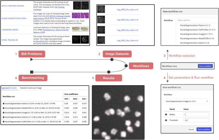
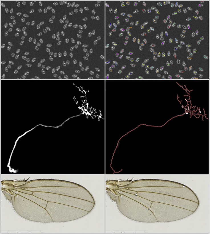
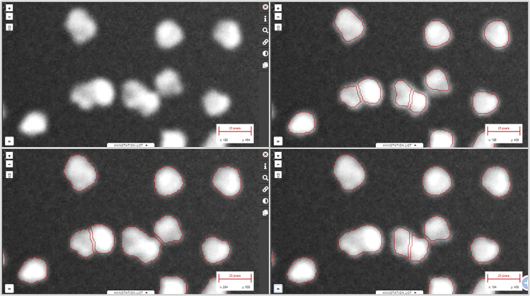
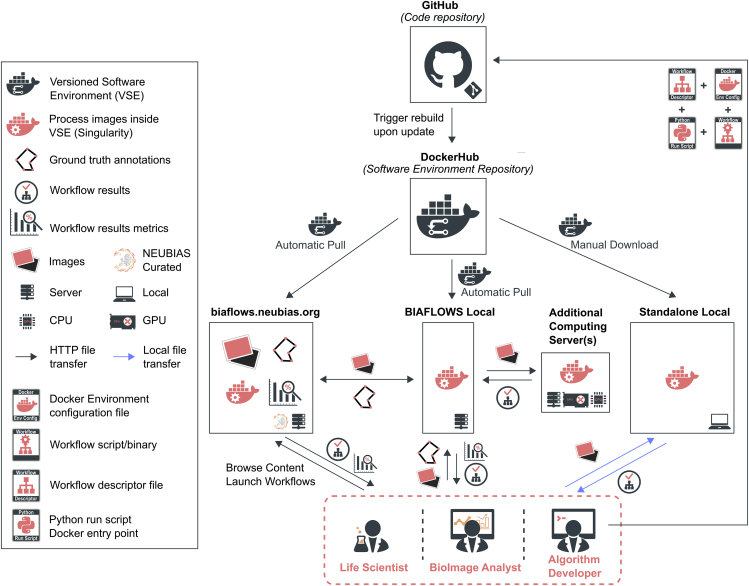
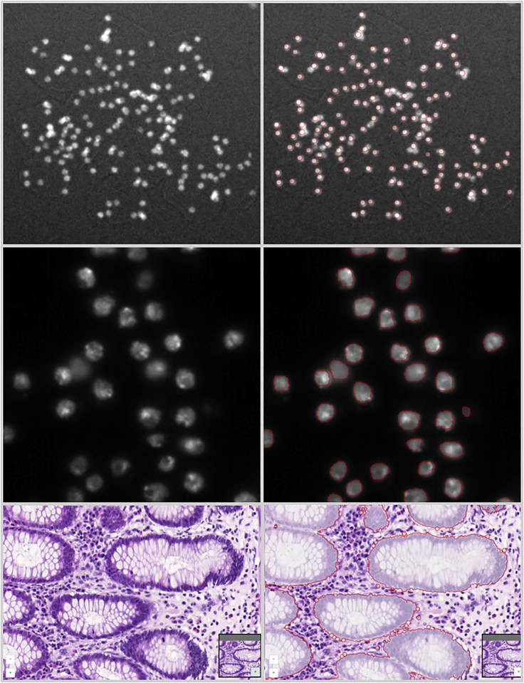
Image analysis is key to extracting quantitative information from scientific microscopy images, but the methods involved are now often so refined that they can no longer be unambiguously described by written protocols. We introduce BIAFLOWS, an open-source web tool enabling to reproducibly deploy and benchmark bioimage analysis workflows coming from any software ecosystem. A curated instance of BIAFLOWS populated with 34 image analysis workflows and 15 microscopy image datasets recapitulating common bioimage analysis problems is available online. The workflows can be launched and assessed remotely by comparing their performance visually and according to standard benchmark metrics. We illustrated these features by comparing seven nuclei segmentation workflows, including deep-learning methods. BIAFLOWS enables to benchmark and share bioimage analysis workflows, hence safeguarding research results and promoting high-quality standards in image analysis. The platform is thoroughly documented and ready to gather annotated microscopy datasets and workflows contributed by the bioimaging community.
图像分析是从科学显微镜图像中提取定量信息的关键,但所涉及的方法现在通常非常精细,以至于书面协议无法再对其进行明确描述。我们引入了BIAFLOWS,这是一个开源网络工具,能够可重复地部署和基准测试来自任何软件生态系统的生物图像分析工作流程。在线提供了一个精心策划的BIAFLOWS实例,其中包含34个图像分析工作流程和15个显微镜图像数据集,概括了常见的生物图像分析问题。通过直观比较工作流程的性能并根据标准基准指标,可以远程启动和评估这些工作流程。我们通过比较包括深度学习方法在内的七种细胞核分割工作流程来说明这些功能。BIAFLOWS能够对生物图像分析工作流程进行基准测试和共享,从而保障研究结果并促进图像分析中的高质量标准。该平台有详尽的文档记录,并准备好收集生物成像社区贡献的带注释的显微镜数据集和工作流程。