Mohammadi Fatemeh, Ashrafi Mahnaz, Zandieh Zahra, Najafi Mohammad, Niknafs Behrooz, Amjadi Fatemeh Sadat, Haghighi Maryam
Student Research Committee, School of Medicine, Iran University of Medical Sciences (IUMS), Tehran, Iran.
Anatomy Department, School of Medicine, Iran University of Medical Sciences (IUMS), Tehran, Iran.
J Reprod Infertil. 2020 Oct-Dec;21(4):259-268. doi: 10.18502/jri.v21i4.4330.
It is demonstrated that optimal preincubation time improves oocyte quality, fertilization potential and developmental rate. This study aimed to evaluate the effect of preincubation time in the simple and myo-inositol supplemented medium on the oocyte quality regarding oxidative stress and mitochondrial alteration.
Cumulus oocyte complexes (COCs) retrieved from superovulated NMRI mice were divided in groups of 0, 4 and 8 preincubation time in the simple and 20 myo-inositol supplemented media. Intracellular reactive oxygen species (HO), glutathione (GSH), mitochondrial membrane potential (MMP), ATP content, and mitochondrial amount were measured and analyzed in experimental groups. One-way ANOVA and Kruskal-Wallis were respectively used for parametric and nonparametric variables. Statistical significance was defined as p<0.05.
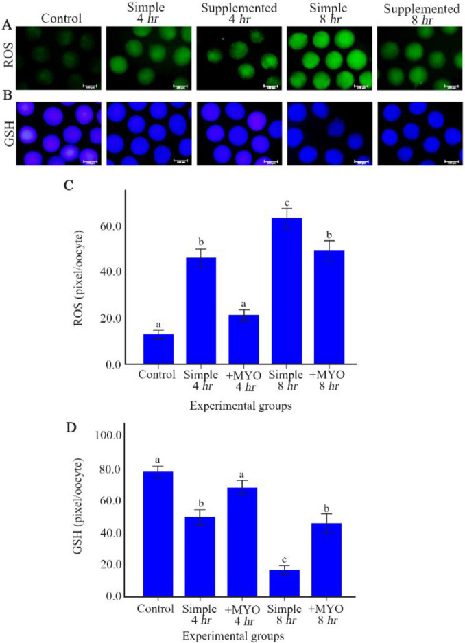
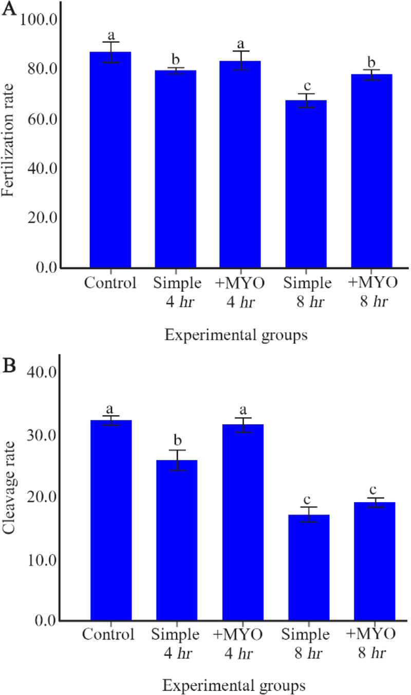
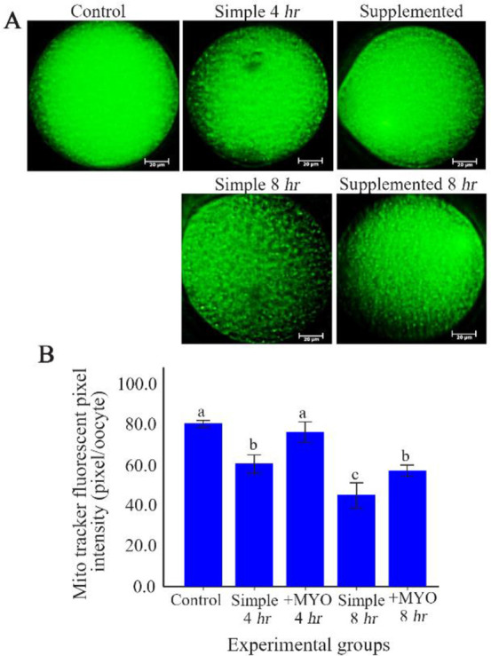
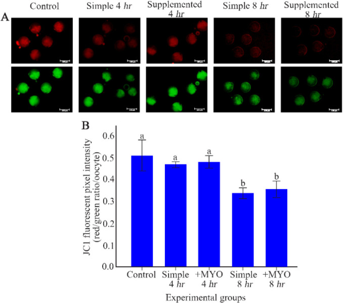
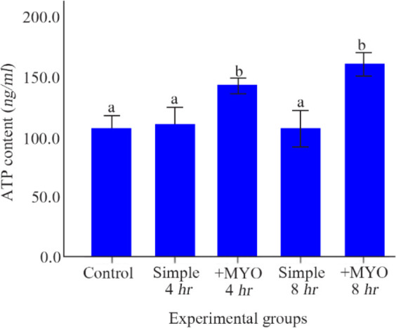
In comparison to control group, variables including ROS, GSH, mitochondrial amount, fertilization and developmental rates were significantly changed after 4 of preincubation in the simple medium, while MMP decreased following 8 of preincubation in the simple medium (p˂0.001). Preincubation of oocytes up to 8 in the simple medium could not decrease ATP content. For both 4 and 8 preincubation times, myo-inositole could decrease HO and increase GSH and MMP levels and consequently could improve fertilization rate compared to oocytes preincubated in the simple culture.
It seems that 4 or more preincubation time can decrease the oocyte quality and lead to reduced oocyte fertilization and developmental potential. Howevere, myo-inositol may prevent oocyte quality reduction and improve fertilization potential in comparision to the equivalent simple groups.
已证明最佳预孵育时间可提高卵母细胞质量、受精潜力和发育率。本研究旨在评估在单纯培养基和添加肌醇的培养基中预孵育时间对卵母细胞质量(涉及氧化应激和线粒体改变)的影响。
从超排卵的NMRI小鼠中获取的卵丘卵母细胞复合体(COCs),在单纯培养基和添加20 μM肌醇的培养基中,根据预孵育时间分为0、4和8小时组。对实验组的细胞内活性氧(ROS)、谷胱甘肽(GSH)、线粒体膜电位(MMP)、ATP含量和线粒体数量进行测量和分析。参数变量和非参数变量分别采用单因素方差分析和Kruskal-Wallis检验。统计学显著性定义为p<0.05。
与对照组相比,在单纯培养基中预孵育4小时后,包括ROS、GSH、线粒体数量、受精率和发育率在内的变量发生了显著变化,而在单纯培养基中预孵育8小时后MMP降低(p˂0.001)。在单纯培养基中对卵母细胞进行长达8小时的预孵育不会降低ATP含量。对于4小时和8小时的预孵育时间,与在单纯培养基中预孵育的卵母细胞相比,肌醇均可降低ROS水平、增加GSH和MMP水平,从而提高受精率。
似乎4小时或更长时间的预孵育会降低卵母细胞质量,并导致卵母细胞受精和发育潜力降低。然而,与同等单纯培养基组相比,肌醇可能会防止卵母细胞质量下降并提高受精潜力。