Department of Psychology, ISEC 672D, Northeastern University, Boston, MA, 02115, USA.
Massachusetts General Hospital, Boston, MA, USA.
Cerebellum. 2021 Jun;20(3):392-401. doi: 10.1007/s12311-020-01213-8. Epub 2020 Nov 18.
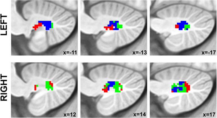
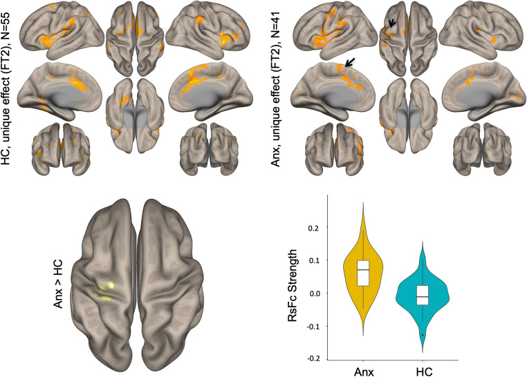
Adolescents with anxiety disorders exhibit excessive emotional and somatic arousal. Neuroimaging studies have shown abnormal cerebral cortical activation and connectivity in this patient population. The specific role of cerebellar output circuitry, specifically the dentate nuclei (DN), in adolescent anxiety disorders remains largely unexplored. Resting-state functional connectivity analyses have parcellated the DN, the major output nuclei of the cerebellum, into three functional territories (FTs) that include default-mode, salience-motor, and visual networks. The objective of this study was to understand whether FTs of the DN are implicated in adolescent anxiety disorders. Forty-one adolescents (mean age 15.19 ± 0.82, 26 females) with one or more anxiety disorders and 55 age- and gender-matched healthy controls completed resting-state fMRI scans and a self-report survey on anxiety symptoms. Seed-to-voxel functional connectivity analyses were performed using the FTs from DN parcellation. Brain connectivity metrics were then correlated with State-Trait Anxiety Inventory (STAI) measures within each group. Adolescents with an anxiety disorder showed significant hyperconnectivity between salience-motor DN FT and cerebral cortical salience-motor regions compared to controls. Salience-motor FT connectivity with cerebral cortical sensorimotor regions was significantly correlated with STAI-trait scores in HC (R = 0.41). Here, we report DN functional connectivity differences in adolescents diagnosed with anxiety, as well as in HC with variable degrees of anxiety traits. These observations highlight the relevance of DN as a potential clinical and sub-clinical marker of anxiety.
焦虑障碍青少年表现出过度的情绪和躯体唤醒。神经影像学研究表明,在这一患者群体中大脑皮质的激活和连通性异常。小脑输出回路,特别是齿状核(DN),在青少年焦虑障碍中的特定作用在很大程度上尚未得到探索。静息态功能连接分析已将 DN 划分为三个功能区(FTs),包括默认模式、突显-运动和视觉网络。本研究的目的是了解 DN 的 FT 是否与青少年焦虑障碍有关。41 名(平均年龄 15.19±0.82,26 名女性)患有一种或多种焦虑障碍的青少年和 55 名年龄和性别匹配的健康对照组完成了静息态 fMRI 扫描和焦虑症状的自我报告调查。使用 DN 分区的 FT 进行种子到体素功能连接分析。然后,在每个组内,将大脑连接度指标与状态-特质焦虑量表(STAI)的测量值相关联。与对照组相比,患有焦虑症的青少年在突显-运动 DN FT 与大脑皮质突显-运动区域之间表现出明显的过度连接。在 HC 中,与大脑皮质感觉运动区域的突显-运动 FT 连接与 STAI-特质评分呈显著相关(R=0.41)。在这里,我们报告了在被诊断为焦虑症的青少年以及在具有不同程度焦虑特征的 HC 中,DN 的功能连接差异。这些观察结果强调了 DN 作为焦虑的潜在临床和亚临床标志物的相关性。