• 文献检索
  • 文档翻译
  • 深度研究
  • 学术资讯
  • Suppr Zotero 插件Zotero 插件
  • 邀请有礼
  • 套餐&价格
  • 历史记录
应用&插件
Suppr Zotero 插件Zotero 插件浏览器插件Mac 客户端Windows 客户端微信小程序
定价
高级版会员购买积分包购买API积分包
服务
文献检索文档翻译深度研究API 文档MCP 服务
关于我们
关于 Suppr公司介绍联系我们用户协议隐私条款
关注我们

Suppr 超能文献

核心技术专利:CN118964589B侵权必究
粤ICP备2023148730 号-1Suppr @ 2026

文献检索

告别复杂PubMed语法,用中文像聊天一样搜索,搜遍4000万医学文献。AI智能推荐,让科研检索更轻松。

立即免费搜索

文件翻译

保留排版,准确专业,支持PDF/Word/PPT等文件格式,支持 12+语言互译。

免费翻译文档

深度研究

AI帮你快速写综述,25分钟生成高质量综述,智能提取关键信息,辅助科研写作。

立即免费体验

存在高对比度时的全波三维逆散射透射超声层析成像。

Full wave 3D inverse scattering transmission ultrasound tomography in the presence of high contrast.

机构信息

QT ultrasound LLC, Novato, USA.

出版信息

Sci Rep. 2020 Nov 19;10(1):20166. doi: 10.1038/s41598-020-76754-3.

DOI:10.1038/s41598-020-76754-3
PMID:33214569
原文链接:https://pmc.ncbi.nlm.nih.gov/articles/PMC7677558/
Abstract

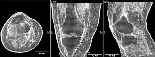
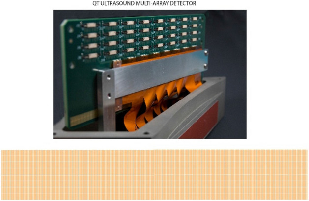
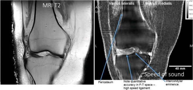
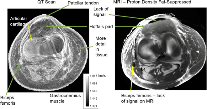
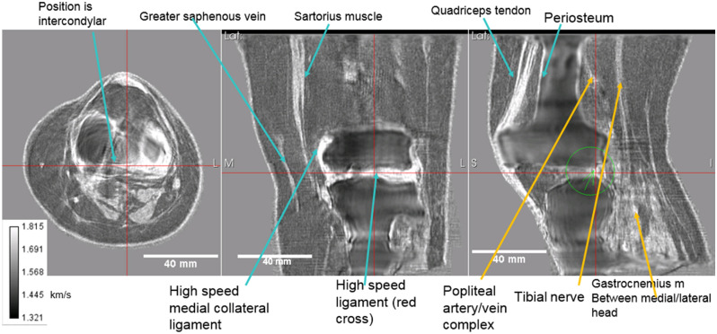
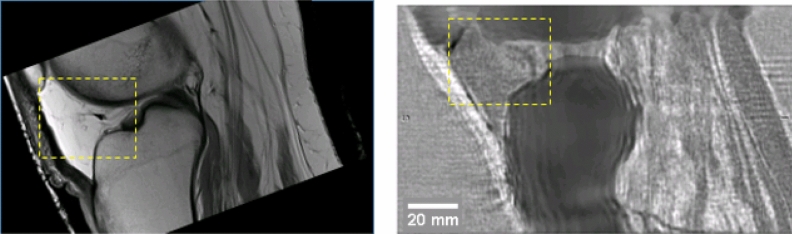
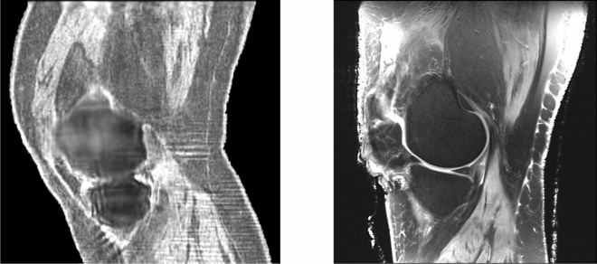
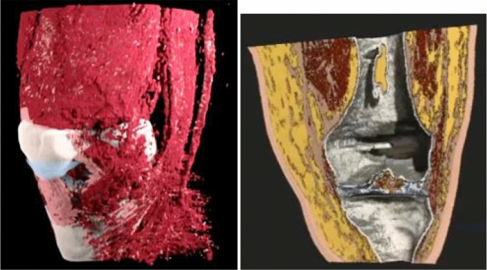
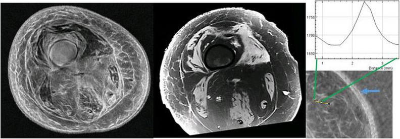
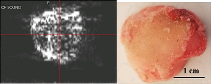
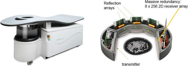
We present here a quantitative ultrasound tomographic method yielding a sub-mm resolution, quantitative 3D representation of tissue characteristics in the presence of high contrast media. This result is a generalization of previous work where high impedance contrast was not present and may provide a clinically and laboratory relevant, relatively inexpensive, high resolution imaging method for imaging in the presence of bone. This allows tumor, muscle, tendon, ligament or cartilage disease monitoring for therapy and general laboratory or clinical settings. The method has proven useful in breast imaging and is generalized here to high-resolution quantitative imaging in the presence of bone. The laboratory data are acquired in ~ 12 min and the reconstruction in ~ 24 min-approximately 200 times faster than previously reported simulations in the literature. Such fast reconstructions with real data require careful calibration, adequate data redundancy from a 2D array of 2048 elements and a paraxial approximation. The imaging results show that tissue surrounding the high impedance region is artifact free and has correct speed of sound at sub-mm resolution.

摘要

我们在这里提出了一种定量超声层析成像方法,可在存在高对比介质的情况下提供具有亚毫米分辨率的组织特性的定量 3D 表示。这一结果是对以前工作的推广,以前的工作中不存在高阻抗对比,并且可能为存在骨骼的成像提供一种具有临床和实验室相关性、相对廉价、高分辨率的成像方法。该方法已在乳腺成像中得到验证,并在此推广到存在骨骼的高分辨率定量成像。实验室数据的采集时间约为 12 分钟,重建时间约为 24 分钟——比文献中以前报道的模拟快约 200 倍。如此快速的重建需要仔细的校准,来自 2048 个元素的二维阵列的充分数据冗余以及傍轴近似。成像结果表明,高阻抗区域周围的组织无伪影,并且在亚毫米分辨率下具有正确的声速。

https://cdn.ncbi.nlm.nih.gov/pmc/blobs/6e37/7677558/3cce991955f4/41598_2020_76754_Fig12_HTML.jpg
https://cdn.ncbi.nlm.nih.gov/pmc/blobs/6e37/7677558/3b8443cf2ea5/41598_2020_76754_Fig1_HTML.jpg
https://cdn.ncbi.nlm.nih.gov/pmc/blobs/6e37/7677558/fb4cc0b8ffe4/41598_2020_76754_Fig2_HTML.jpg
https://cdn.ncbi.nlm.nih.gov/pmc/blobs/6e37/7677558/0fe3d7d997f6/41598_2020_76754_Fig3_HTML.jpg
https://cdn.ncbi.nlm.nih.gov/pmc/blobs/6e37/7677558/2fea5ae42099/41598_2020_76754_Fig4_HTML.jpg
https://cdn.ncbi.nlm.nih.gov/pmc/blobs/6e37/7677558/5ad4e1445f6a/41598_2020_76754_Fig5_HTML.jpg
https://cdn.ncbi.nlm.nih.gov/pmc/blobs/6e37/7677558/f087e7b31923/41598_2020_76754_Fig6_HTML.jpg
https://cdn.ncbi.nlm.nih.gov/pmc/blobs/6e37/7677558/4d65c9a485b7/41598_2020_76754_Fig7_HTML.jpg
https://cdn.ncbi.nlm.nih.gov/pmc/blobs/6e37/7677558/b2a276e29f40/41598_2020_76754_Fig8_HTML.jpg
https://cdn.ncbi.nlm.nih.gov/pmc/blobs/6e37/7677558/32c9c5d6474f/41598_2020_76754_Fig9_HTML.jpg
https://cdn.ncbi.nlm.nih.gov/pmc/blobs/6e37/7677558/bd650a096b04/41598_2020_76754_Fig10_HTML.jpg
https://cdn.ncbi.nlm.nih.gov/pmc/blobs/6e37/7677558/4b7e716e0fbe/41598_2020_76754_Fig11_HTML.jpg
https://cdn.ncbi.nlm.nih.gov/pmc/blobs/6e37/7677558/3cce991955f4/41598_2020_76754_Fig12_HTML.jpg

相似文献

1
Full wave 3D inverse scattering transmission ultrasound tomography in the presence of high contrast.存在高对比度时的全波三维逆散射透射超声层析成像。
Sci Rep. 2020 Nov 19;10(1):20166. doi: 10.1038/s41598-020-76754-3.
2
Low frequency 3D transmission ultrasound tomography: technical details and clinical implications.低频三维传输超声断层成像:技术细节与临床应用
Z Med Phys. 2023 Aug;33(3):427-443. doi: 10.1016/j.zemedi.2023.04.006. Epub 2023 Jun 7.
3
Cancellous and cortical bone imaging by reflected tomography.通过反射断层扫描对松质骨和皮质骨进行成像。
Ultrason Imaging. 2001 Jan;23(1):55-70. doi: 10.1177/016173460102300104.
4
Three-dimensional simulation of ultrasound propagation through trabecular bone structures measured by synchrotron microtomography.通过同步加速器显微断层扫描测量的小梁骨结构中超声传播的三维模拟。
Phys Med Biol. 2005 Dec 7;50(23):5545-56. doi: 10.1088/0031-9155/50/23/009. Epub 2005 Nov 8.
5
Two-wave propagation imaging to evaluate the structure of cancellous bone.双波传播成像评估松质骨结构。
IEEE Trans Ultrason Ferroelectr Freq Control. 2012 Jun;59(6):1160-6. doi: 10.1109/TUFFC.2012.2306.
6
Deep convolutional neural network for segmentation of knee joint anatomy.深度卷积神经网络用于膝关节解剖结构的分割。
Magn Reson Med. 2018 Dec;80(6):2759-2770. doi: 10.1002/mrm.27229. Epub 2018 May 17.
7
Tomographic reconstruction of three-dimensional volumes using the distorted born iterative method.基于扭曲玻恩迭代法的三维容积断层重建。
IEEE Trans Med Imaging. 2009 Oct;28(10):1643-53. doi: 10.1109/TMI.2009.2026274. Epub 2009 Jun 30.
8
Model-based ultrasound tomography: tissue phantom experiments.基于模型的超声层析成像:组织体模实验
Med Phys. 2005 Aug;32(8):2659-64. doi: 10.1118/1.1990287.
9
Numerical simulation of wave propagation in cancellous bone.松质骨中波传播的数值模拟。
Ultrasonics. 2006 Dec 22;44 Suppl 1:e239-43. doi: 10.1016/j.ultras.2006.06.042. Epub 2006 Jun 30.
10
Quantitative transmission ultrasound tomography: Imaging and performance characteristics.定量超声层析成像:成像和性能特征。
Med Phys. 2018 Jul;45(7):3063-3075. doi: 10.1002/mp.12957. Epub 2018 May 28.

引用本文的文献

1
Inverse-scattering in biological samples via beam-propagation.通过光束传播对生物样本进行逆散射。
bioRxiv. 2025 Aug 22:2025.08.17.670744. doi: 10.1101/2025.08.17.670744.
2
Angular spatial compounding of diffraction corrected images improves ultrasound attenuation measurements.衍射校正图像的角向空间复合改善了超声衰减测量。
J Acoust Soc Am. 2025 Mar 1;157(3):1638-1649. doi: 10.1121/10.0036124.
3
Breast Glandular and Ductal Volume Changes during the Menstrual Cycle: A Study in 48 Breasts Using Ultralow-Frequency Transmitted Ultrasound Tomography/Volography.

本文引用的文献

1
Emerging methods in radiology.新兴的放射学方法。
Radiologe. 2020 Nov;60(Suppl 1):41-53. doi: 10.1007/s00117-020-00696-0.
2
Full-waveform inversion imaging of the human brain.人类大脑的全波形反演成像。
NPJ Digit Med. 2020 Mar 6;3:28. doi: 10.1038/s41746-020-0240-8. eCollection 2020.
3
Contrast-Enhanced Ultrasound Quantification: From Kinetic Modeling to Machine Learning.对比增强超声定量:从动力学建模到机器学习。
经超低频发射超声断层摄影/体层摄影术研究 48 例乳房,了解月经周期中乳腺腺泡和导管体积的变化。
Tomography. 2024 May 19;10(5):789-805. doi: 10.3390/tomography10050060.
4
Spectral-based Quantitative Ultrasound Imaging Processing Techniques: Comparisons of RF Versus IQ Approaches.基于光谱的定量超声成像处理技术:RF 与 IQ 方法的比较。
Ultrason Imaging. 2024 Mar;46(2):75-89. doi: 10.1177/01617346231226224. Epub 2024 Feb 6.
5
In vivo noninvasive systemic myography of acute systemic vasoactivity in female pregnant mice.体内无创性系统性血管活性研究在雌性怀孕小鼠急性系统性血管活性中的应用。
Nat Commun. 2023 Oct 9;14(1):6286. doi: 10.1038/s41467-023-42041-8.
6
A Forward Model Incorporating Elevation-Focused Transducer Properties for 3-D Full-Waveform Inversion in Ultrasound Computed Tomography.一种结合聚焦于高程的换能器特性的正向模型,用于超声计算机断层扫描中的三维全波形反演。
IEEE Trans Ultrason Ferroelectr Freq Control. 2023 Oct;70(10):1339-1354. doi: 10.1109/TUFFC.2023.3313549. Epub 2023 Oct 17.
7
Full Wave Inversion and Inverse Scattering in Ultrasound Tomography/Volography.超声层析成像/容积成像中的全波反演和逆散射。
Adv Exp Med Biol. 2023;1403:201-237. doi: 10.1007/978-3-031-21987-0_10.
8
Low frequency 3D transmission ultrasound tomography: technical details and clinical implications.低频三维传输超声断层成像:技术细节与临床应用
Z Med Phys. 2023 Aug;33(3):427-443. doi: 10.1016/j.zemedi.2023.04.006. Epub 2023 Jun 7.
9
An Experimental Ultrasound System for Qualitative Tomographic Imaging.一种用于定性断层成像的实验性超声系统。
Sensors (Basel). 2022 Oct 14;22(20):7802. doi: 10.3390/s22207802.
10
3-D Stochastic Numerical Breast Phantoms for Enabling Virtual Imaging Trials of Ultrasound Computed Tomography.用于实现超声计算机断层成像虚拟成像试验的三维随机乳房体模。
IEEE Trans Ultrason Ferroelectr Freq Control. 2022 Jan;69(1):135-146. doi: 10.1109/TUFFC.2021.3112544. Epub 2021 Dec 31.
Ultrasound Med Biol. 2020 Mar;46(3):518-543. doi: 10.1016/j.ultrasmedbio.2019.11.008. Epub 2020 Jan 8.
4
Quantitative Assessment of Breast Density: Transmission Ultrasound is Comparable to Mammography with Tomosynthesis.乳腺密度定量评估:超声传输与断层合成钼靶 X 线摄影相当。
Cancer Prev Res (Phila). 2019 Dec;12(12):871-876. doi: 10.1158/1940-6207.CAPR-19-0268. Epub 2019 Oct 23.
5
Preoperative Ultrasound Radiomics Signatures for Noninvasive Evaluation of Biological Characteristics of Intrahepatic Cholangiocarcinoma.术前超声放射组学特征用于无创评估肝内胆管细胞癌的生物学特征。
Acad Radiol. 2020 Jun;27(6):785-797. doi: 10.1016/j.acra.2019.07.029. Epub 2019 Sep 5.
6
iPSCs: A powerful tool for skeletal muscle tissue engineering.iPSCs:骨骼肌组织工程的有力工具。
J Cell Mol Med. 2019 Jun;23(6):3784-3794. doi: 10.1111/jcmm.14292. Epub 2019 Apr 1.
7
Quantitative assessment of breast density using transmission ultrasound tomography.应用透射超声层析成像技术对乳腺密度进行定量评估。
Med Phys. 2019 Jun;46(6):2610-2620. doi: 10.1002/mp.13503. Epub 2019 Apr 22.
8
Radiomics: the facts and the challenges of image analysis.放射组学:图像分析的现状与挑战
Eur Radiol Exp. 2018 Nov 14;2(1):36. doi: 10.1186/s41747-018-0068-z.
9
Decellularized Tissue for Muscle Regeneration.脱细胞组织用于肌肉再生。
Int J Mol Sci. 2018 Aug 14;19(8):2392. doi: 10.3390/ijms19082392.
10
Radiomic analysis of contrast-enhanced ultrasound data.对比增强超声数据的放射组学分析。
Sci Rep. 2018 Jul 27;8(1):11359. doi: 10.1038/s41598-018-29653-7.